“The Best” Disease You’ve Never Heard Of: A Tale of Two Patients

Juan Ding, OD, PhD

When people think of inherited eye diseases, they usually imagine problems that show up in babies or childhood, with both eyes blind. But what if I told you the same rare eye condition can show up in kids as well as the elderly — and affecting two eyes differently?

Recently, I saw two patients who were diagnosed with a condition called Best vitelliform macular dystrophy, more commonly known as Best disease. Despite the name, it’s not exactly “the best” news to receive — though, thankfully, it tends to progress slowly and isn’t typically associated with total blindness.

Let’s start with the little girl. This is a sweet 6-year-old girl who failed school vision screening and referred to our clinic. She reports that she sees everything well, but parents note that she stays close to the TV to watch. After a detailed eye exam and specialized imaging, I diagnosed her with Best disease. She was otherwise completely healthy, with no complaints — though her vision is 20/60 in both eyes.

Interestingly, even though both eyes are not seeing well, they do show very different appearances in photos. Below is the color photo and cross-sectional view of her retina (Figure 1). Her right eye is at the ‘egg yolk’ stage, while the left eye has already progressed to the late, or advanced stage of macular atrophy. My heart goes out to the family. She is so young, and yet already at the late stage of this disease. Fortunately her vision is not too bad. And that is a common feature of this condition, that vision is typically only moderately impaired even in the late stage.

Figure 1. Photos of retina in a 6 year old girl with Best disease. The two columns represent right and left eye as labeled. A, color photo of the retina with subtle changes in macular appearance. B, autofluorescent black and white photo of the macula area. C, OCT cross-section photo of the macula in both eyes. Yellow arrow points to the ‘egg yolk’ in the macula of the right eye; white arrow points to a thin and atrophied macula in the left eye.

Then came the man in his sixties a week later. The two patients are not related. Just a coincidence that I would see two cases of a rare eye condition in the span of a week in a primary care setting. He started noticing blurry vision in the right eye when he was 50. He came to see us in 2021 but then lost to follow up. It gradually got worse, and now his right eye sees 20/40. His left eye sees 20/20 and no change over the years. Again, imaging confirmed the diagnosis. What is interesting is that you can see that over the 4 years, the right eye has progressed from the ‘egg yolk’ stage to ‘atrophy’ stage; while the left eye has little progression with still early or small ‘egg yolk’.

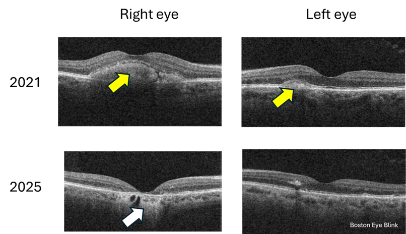
Figure 2. Cross-section macular photo of a 60 year old man in 2021 and 2025. Yellow arrow points to the ‘egg yolk’; white arrow points to atrophied retina.

So what exactly is Best disease?

This condition is so named not because it’s the best disease a person can have, it’s named after Dr Franz Best, a German ophthalmologist, who described the first pedigree in 1905.

Best disease is a rare genetic condition that affects the macula, the central part of the retina responsible for sharp, detailed vision. It’s caused by mutations in a gene called BEST1 [1], which affects a layer of cells beneath the retina called the retinal pigment epithelium (RPE). This layer helps keep the retina healthy and functioning properly. The prevalence is about 1 in 20,000 [2], so very rare.

The condition often runs in families and usually appears in childhood or adolescence, though some people — like my older patient — might not show symptoms until later in life. However, the adult onset Best disease may also be caused by mutations in genes other than BEST1 [1]. The onset of age ranges from 3-15 years of age [3]. It typically affects both eyes, though in some cases the two eyes show different rate of progression, which happens to be in both my patients.

Best disease is inherited in an autosomal dominant manner, meaning if there is one faulty gene, the disease will manifest. Therefore if one person has the disease, some of their family members are likely to have it too. But the degree and extensiveness of the disease can be quite variable among individuals [1], sometimes even with the condition, the person may not have any symptoms.

Interestingly, for both of my patients, they report no knowledge of any family history of low vision or blindness. I highly recommend the direct relatives to have a comprehensive eye exam with retinal imaging – the most important being the fundus imaging and OCT which can show cross section of the retina tissue. This technology can show the most striking feature, the yellowish lesion in the macula that looks a bit like an egg yolk — which is why doctors often call it a “vitelliform lesion.” You can see these lesions in the girl and the man in Figures 1 and 2 above. Over time, this lesion can change shape, break up, or leak fluid, leading to vision changes. In late stages, the macula becomes atrophied, meaning the tissue dies and thins out.

Can it be treated?

There’s no cure for Best disease, but the good news is that many people maintain useful vision throughout their lives. Regular monitoring is key to catching any complications early, especially if fluid builds up or if abnormal blood vessels form — in which case treatment might involve medications or laser therapy.

Recently, people are attempting gene therapy of this condition in dog models [4]. Hopefully gene therapy becomes available in the future in humans affected by this condition.

Why does this matter?

These two patients — so different in age and life stage — are a reminder that eye disease doesn’t always follow predictable rules. For example, Best disease usually affects both eyes, but these two patients both show remarkable asymmetry in stages of the disease. Best disease progresses slowly, and while it is true for the elder man, for the little girl, one eye is already at advanced stage. Given her young age, I imagine the progression has been fast.

And while it may not be “the best” diagnosis to receive, with proper care and awareness, patients can still live full, visually rich lives.

References and extended readings

[1] https://eyewiki.org/Best_Disease_and_Bestrophinopathies

[2] Tripathy K, Salini B. Best Disease. [Updated 2023 Aug 25]. In: StatPearls [Internet]. Treasure Island (FL): StatPearls Publishing; 2025 Jan-. Available from: https://www.ncbi.nlm.nih.gov/books/NBK537290/

[3] https://emedicine.medscape.com/article/1227128-overview#:~:text=gender%20predilection%20exists.-,Age,occurs%20after%20age%2040%20years.

[4] Amato A, Wongchaisuwat N, Lamborn A, Schmidt R, Everett L, Yang P, Pennesi ME. Gene therapy in bestrophinopathies: Insights from preclinical studies in preparation for clinical trials. Saudi J Ophthalmol. 2023 Dec 1;37(4):287-295. doi: 10.4103/sjopt.sjopt_175_23. PMID: 38155675; PMCID: PMC10752275.

My Unexpected Spin: Living Through BPPV

by Juan Ding, OD, PhD

I am not an ENT doctor or vestibular specialist, however, I will write about my recent personal experience on vertigo as a patient and hope it’s helpful for others who suffer from this condition.

Through my optometry practice, I frequently encounter patients referred by PCPs or ENT doctors for vertigo. It’s certainly not primarily an eye disease, but often doctors want to rule out eye conditions that mimic vertigo. Many of these patients have Benign Paroxysmal Positional Vertigo, or BPPV. They would describe to me how the room spins when it attacks. I often listen with great sympathy, and never imagined I’d wake up one morning and the room would be spinning. One turn of my head and suddenly, my world tilted, my stomach lurched, and I was slammed with a wave of nausea. I had no fever, no ear infection, no warning. Just intense vertigo.

The Onset

It was 4 or 5 AM, I woke up to go to the bathroom. This was the last day of our vacation in Florida, and I expected some nice relaxation before the flight later in the afternoon. After several days of the excitement and exhaustion at Disney and Universal Studios, I really looked forward to a peaceful day and return home.

However, as soon as I got up, I noticed the world was spinning. I managed to use the bathroom, and returned to bed. After laying down, spinning again. I immediately realized I was having BPPV, my very first episode. It was positional and lasted only a minute. If I tilted or turned my head a certain way—especially lying down or rolling over in bed—it would trigger another episode.

The Culprit: Inner Ear Crystals

Even though I knew about the condition, and could self-diagnose quickly, I had no idea how to treat it. I learned that BPPV is caused by tiny calcium crystals (otoconia, Figure below) that normally sit in a gel-like structure in your inner ear, helping your brain sense gravity and balance. Sometimes, these crystals dislodge and drift into the semicircular canals where they don’t belong, triggering false signals when you move your head.

Thanks to youtube and ChatGPT, I learned that the treatment is Epley Maneuver, a series of head movements designed to guide the crystals back where they belong. It makes sense, and it looks simple enough.

My attempts with the Epley Maneuver

Various youtube videos, many by vestibular physical therapists, demonstrate how to do this yourself at home. All you need is a bed and a pillow. I thought, I could do this. The first time I tried—I didn’t do it quite right. I was not tilting my head backward enough. And I had to check the video half way though the movement to see what happened next.

The second and third tries improved, but after each attempt, I was left extremely nauseated. I was knocked down and could not get up, dare not get up, I felt extremely sick and depressed. I must be doing something wrong, how come all the comments under those videos were from grateful patients that tried it once and were cured?

But here’s the thing that I later found out: you don’t always feel better immediately. In fact, the maneuver itself can make you feel worse before it makes you better. The key indicator of success? A reduction in spinning when you repeat the same positions. Dizziness and lightheadedness may linger, but the violent spinning should ease up.

That day I became a robot. I moved extremely slowly with absolutely no head movement for fear of another attack. It was a miracle that I got on the plane and returned home. In my mind, I would be calling 911 at some point and ended up in a hospital, not home.

I later learned that many people do go to the emergency room for this. Imagine the panic you have when you wake up with the world spinning and you feeling dizzy and sick. Every head movement is a potential trigger and it’s easy to think that something is seriously wrong. Even when I know the diagnosis, I feel desperate after a few attempts of treatment and not getting the resolution. There is nothing benign about this supposedly benign condition. You don’t want to live with vertigo, the nausea and the sickness it brings about.

Living in fear

In the days that followed, I was cautious. I avoided quick movements, slept with my head elevated on two pillows, and limited head-turning. I noticed a pattern—symptoms were worse in the morning and improved as the day went on. This made sense, as lying down at night likely allowed crystals to shift again.

I called multiple vestibular PT offices, unfortunately everyone was closed on weekends. I was ready to ask for emergency sick leave should I still be sick on Monday. I thought that I would only be able to get better after seeing a specialist who does this Epley maneuver properly on me.

I browsed through scores of videos on perfecting the Epley maneuver. I got really good at it when I would do it again and again in my mind, though I was afraid to actually try it, because I knew there would be more sickness afterwards.

I learned that in China and Europe there are machines that do this, you just need to be strapped on it and it will do it and you will be cured. I wish this were available here.

Final recovery

On Sunday morning, I felt better and decided to give the Epley maneuver one last go. If I should fail again, I would see PT and they could fix it. I prayed that it would work.

And it worked, but also didn’t. It didn’t, because despite doing every step correctly, there was no vertigo this time. Which means that the crystals did not shift. It worked, because it actually already worked 2 days before. The reason that I did not have any vertigo this time was because the crystals already were repositioned from my previous attempts. I didn’t know it had worked because I continued to have intense dizziness and nausea for the two days after, but in reality it was the recovery of the vestibular system after a brutal crash. Yes it did take that long for me. And all this time that I did not have vertigo, I thought was due to me being a proper robot, but it’s because the runaway crystals had returned home.

In the days that followed, I continued to get a little better each day. I was never this grateful when I could bend down to pick up a toy from the floor without feeling sick.

After a total of 6 days, I returned to my baseline.

Risk Factors for BPPV

So why me? And why you?

BPPV can strike anyone, but certain factors raise the risk [1]:

  • Age: It’s more common after 50. This is because the otoconia become weaker as we get older. A similar analogy is the vitreous gel becomes weaker during aging.
  • Head injury: Even minor trauma can dislodge otoconia
  • Inner ear disorders. Infection and inflammation of the inner ear may loosen the otoconia
  • Prolonged bed rest. When laying down, especially on our sides, the otoconia are more easily translocating into the semicircular canals due to gravity.
  • Migraine history. A theory says that poor circulation of the head and the inner ear may predispose an individual to migraine and BPPV, respectively.
  • Vitamin D deficiency. Otoconia are calcium crystals and their metabolism relies on vitamin D.
  • Dehydration. The inner ear fluid is quite delicate and can be affected by dehydration.

In my case, I do have a history of migraine. I just went through 6 months of New England winter and would be properly vitamin D deficient. I went on a few mild rides at Disney and Universal studios, which could have loosened up those crystals in the inner ear. And I was definitely dehydrated under the glowing Florida sun.

How to Prevent Recurrence

My biggest fear now is a recurrence. I don’t want to live though it again, ever. Research shows that up to 37% people experience a recurrent episode [2].

Although BPPV can return, there are a few measures that may help reduce the risk:

  • Avoid sudden head movements, especially when lying down or getting up
  • Sleep with your head slightly elevated
  • Change positions slowly, especially after waking
  • Stay hydrated and keep your vitamin D levels up
  • Regular neck and vestibular exercises (once you’ve fully recovered)
  • Avoid sleeping on the “trigger” side if you know which one it is

I would be doing all of the above for the rest of my life. Plus, no rollercoaster rides ever again.

Final Thoughts

BPPV is common, yet many people haven’t heard of it until they’re hit with a frightening episode of vertigo. The good news is—it’s treatable. And for most people, repositioning maneuvers work well.

For me, it’s been a journey of listening to my body, slowing down, and learning the science behind balance. If you’ve ever felt like your world is spinning out of control—literally—you’re not alone.

References

[1] Chen J, Zhao W, Yue X, Zhang P. Risk Factors for the Occurrence of Benign Paroxysmal Positional Vertigo: A Systematic Review and Meta-Analysis. Front Neurol. 2020 Jun 23;11:506. doi: 10.3389/fneur.2020.00506. PMID: 32655479; PMCID: PMC7324663.

[2] Sakaida M, Takeuchi K, Ishinaga H, Adachi M, Majima Y. Long-term outcome of benign paroxysmal positional vertigo. Neurology. 2003 May 13;60(9):1532-4. doi: 10.1212/01.wnl.0000061477.03862.4d. PMID: 12743247.