A guide to phakic intraocular lenses (pIOLs) for myopia correction

Phakic IOLs are small lenses implanted in the eye to correct refractive error. So far, FDA has approved two lenses to be used for correcting myopia up to -20.00 diopters (D) with astigmatism up to 2.50 D for people 21-45 years of age. These are alternatives to corneal refractive surgery such as LASIK, especially for high myopia, because too much corneal tissue needs to be removed in high myopia in LASIK and it becomes unsafe. As a comparison, LASIK is FDA approved for myopia up to -11.00 D. Click here to read my post on LASIK.

Two implantable lenses approved by FDA

  • The Visian ICL (Implantable Collamer Lens), approved in 2005, is placed behind the iris and in front of the natural lens. It is invisible to the naked eye. The Visian ICL is made of a soft, biocompatible collagen copolymer, and can be folded during implantation, requiring only a small incision of 3 mm.
  • The Verisyse (branded as Artisan in Europe), approved in 2004, is placed in front of the iris, also called an iris-claw lens. The Verisyse lens is made of a rigid plastic, and because it cannot be folded, it requires a larger incision of 6 mm. If you look very carefully, you may see the lens with naked eyes.

Are you a good candidate for pIOL implant?

As mentioned earlier, patients with high myopia outside the range for LASIK can have pIOL implant to correct myopia. However, you still need to meet certain requirements to be a good candidate. Below are some of the important factors to consider.

  1. pIOL is approved for myopia up to -20.00 D, not higher
  2. Two important parameters of the eye need to be determined: the anterior chamber of your eye has to be deep enough; and your corneal endothelium needs to be healthy. This is because the implanted lens needs to have enough room in the eye, otherwise it may block the drainage system of the eye and causes increased eye pressure and glaucoma. In addition, the surgery itself and the lens often cause loss of corneal endothelial cells, as will be discussed further later, therefore a healthy corneal endothelium is also a pre-requisite.
  3. You must have had stable glass prescription for at least a year.
  4. Eye conditions such as cataracts, glaucoma and untreated eye infections will prevent you from being a good candidate.
  5. Systemic contraindications include Sjogren’s syndrome, rheumatoid arthritis, diabetes, HIV and AIDS, and certain medications such as steroids and immunosuppressants may interfere with healing and final outcomes.

What does the surgery involve?

Prior to the surgery, a procedure called laser peripheral iridotomy (LPI) will be performed on the eye that will receive the implant. This procedure makes a hole on the periphery of your iris to prevent the eye pressure from going up during and post-surgery. LPI can be done a week before or on the same day of the surgery. A newer model lens available in Europe and China but not in the US at the moment, Visian ICL V4c, which has a 0.36 mm diameter hole in the center of the lens, allows fluid exchange without the need of this extra procedure.

The surgery itself takes 10 to 30 minutes and is performed on an outpatient basis. Numbing eyes drops are applied, then small incisions made on the cornea to allow the lens to be inserted into the eye. Here is a demonstration video showing the procedure of Visian ICL: https://www.youtube.com/watch?v=wlaAYBefNTo; and for Verisyse lens: https://www.youtube.com/watch?v=Y4GUpMAHA9s.

After the surgery, you will be given antibiotic and anti-inflammatory eye drops to prevent infection and inflammation.

Most patients will notice clear vision shortly after the surgery and their vision stabilizes within a week, but for some patients it may take a few weeks. There is minimum pain although foreign body sensation can be common after the surgery. Most people can resume work and normal daily activities within a few days.

Safety and Efficacy of pIOLs

Phakic IOL implantation is considered to be safe. However, this is still an open-eye surgery, so risks including retinal detachment and endophthalmitis (infection of the entire eye) due to the surgery exist. Generally, the surgery risk is less than that of cataract surgery, but more than LASIK where the eye remains a closed system.

The two types of pIOLs showed equal and comparable safety, predictability, and efficacy [1].

The accuracy of the optics is decent. For Visian ICL, in 41 eyes of 41 patients with myopic refractive errors of -4.00 to -15.25 D, at 8 years, 68.3% and 85.4% of the eyes were within 0.5 and 1.0 D, respectively, of the targeted correction [2].

The vision post-surgery is also good. In FDA trials, the Verisyse have been shown to have 20/40 uncorrected visual acuity (UCVA) or better in 84% of patients after three years and Visian ICLs have been shown to have an UCVA of 20/40 or better in 81% of patients after 3 years [3]. A different study found 60.5% eyes with UCVA 20/20 or better [4].

Considering that this patient population typically have myopia -10.00 D and more, it is not hard to imagine that patient satisfaction is high overall.

So in summary, pIOLs to correct myopia show good safety and efficacy. It may not be completely fair to compare the UCVA post pICLs vs LASIK, as patients in pICLs typically are more myopic than those in LASIK, and we know that some high myopes may not achieve best corrected VA of 20/20 due to myopic retinal changes.

Just by looking at the numbers, the percentage of eyes within targeted correction appears to be lower than that of LASIK (98.6% of eyes reach refraction within 1.00 D of target, and 90.9% of eyes reach within 0.50 D of target refraction). Given that we are comparing two types of refractive surgeries in two different patient populations, one cannot make a conclusion that LASIK achieves better correction than ICL; but as a patient, you can roughly estimate your chance of getting the ideal correction if you are a candidate for LASIK or pIOL.

Complications of ICL

There are several complications, most common are cataract and corneal endothelial cell loss. Verisyse lenses and Visian ICLs are similar, since Visian ICLs are better studied, I will use ICLs as an example for this section. Below are data summarized by a recent meta-analysis [5] unless otherwise indicated.

ICL replacement

The surgery is reversible, meaning that the implant can be taken out or replaced if it is not ideal. Still, no one likes to have a second surgery. Fortunately, only 1.0-2.6% of cases require a replacement of ICL. The common causes for replacement include too long or too short of a distance between the implanted lens and the natural lens.

Cataract

ICL induces a specific cataract called anterior subcapsular cataract (ASC) due to close proximity of the implant to the front part of the natural lens. Based on 8 studies, it is estimated that the incidence of ASC due to ICL implant ranges from 1.1% to 5.9%, and the incidence of ASC cataracts requiring surgery ranges from 0% to 1.8%.

Another review looking at 2592 eyes showed ASC in 5.2% cases, of these, 43.4% were reported within 1 year, 15.4% between 1 and 3 years, and 35.3% ≥ 3 years after ICL implantation [6].

Even though the cataract incidence due to implants remains low, it has been found that for older patients (>40 years of age) and higher myopia (-12.00 D or higher), the risk of cataract were much increased, as high as 28% at 6 year follow up.

High myopia itself is a risk factor for ASC post ICL implant. While clinically significant cataracts occurred in seven (6.6%) of 106 eyes with preoperative myopia of −12.00 D or higher, none occurred in the 420 eyes with preoperative myopia lower than −12.00 D.

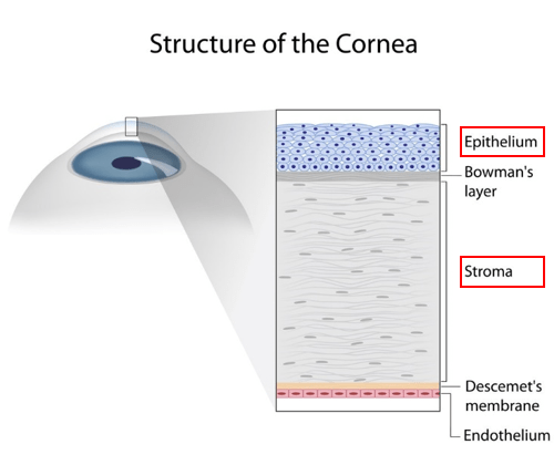
Corneal endothelial cell loss

Corneal endothelial cells are important in keeping the cornea clear. These cells do not regenerate and we lose about 0.4% of them every year as a result of normal aging [7]. ICL implantation accelerates the endothelial cell loss to about 7.5% loss at 5 year follow up. Fortunately, this stabilizes after 2-3 years.

Glaucoma

Lens implantation may cause release of pigment from the iris, blocking the drainage system of the eye, thereby increasing eye pressure, leading to glaucoma. Only two  eyes  out  of  526  (0.4%)  were found to have increased  eye pressure requiring  treatment  at  3  years  post op.

Iris atrophy and pupil distortion

Since the surgery involves placing a lens close to the iris at the center where the pupil is, risk for iris atrophy or pupil distortion exist. In following up with 993 eyes undergoing ICL implantation from 1996 to 2008, iris atrophy and pupil abnormality were found in 0.2% of cases [8]. Higher incidence of iris atrophy was found in the iris-claw type Verisyse lens, 11.8% (11 out of 93 eyes) [9]. This may be skewed from being a study with smaller number of patients, but it is also not a surprise, as the Verisyse lens is fixed on the front of the iris.

So, pIOLs or LASIK?

First of all, remember that all surgeries carry risks, and that wearing spectacles is safe and involves no manipulation in your eyes.

However, if refractive surgery is something you want to go for, make sure you know the indications, benefits, risks and complications associated with each. LASIK is approved for myopia up to -11.00 D, and appropriate corneal thickness is required. Common complications include dry eye, and rare but more serious complication such as corneal ectasia can be detrimental to vision.

pIOLs are better for high myopia up to -20.00 D, does not require a thick cornea but requires healthy corneal endothelial cells and a deep anterior chamber. Complications include cataract and corneal endothelial cell loss; and, as a more invasive procedure, retinal detachment and eye infections can happen though very rare. Caution to older patients and those with higher myopia, as risk of cataract goes up significantly in these patient populations.

In terms of efficacy, both are quite good with high patient satisfaction, though LASIK achieving slightly better correction to target.

In terms of long-term safety, LASIK has been approved in the US for over 20 years whereas pIOLs for 12 years, both have been relatively safe with small percentages of complications as discussed in this article and before.

Lastly both procedures are elective and not covered by insurance; pIOL costs about $4,000 per eye, whereas LASIK about $2,000 per eye in the US.

If you are interested in refractive surgeries, make sure you go for complete eye exams and rule out eye and systemic conditions that contraindicate these surgeries. Eye doctors will recommend the appropriate procedure based on your specific eye conditions and systemic health. Make sure you follow doctors’ instructions for pre and post op care to prevent unnecessary complications.


References


  1. Hassaballa, M.A. and T.A. Macky, Phakic intraocular lenses outcomes and complications: Artisan vs Visian ICL. Eye (Lond), 2011. 25(10): p. 1365-70.
  2. Igarashi, A., K. Shimizu, and K. Kamiya, Eight-year follow-up of posterior chamber phakic intraocular lens implantation for moderate to high myopia. American Journal of Ophthalmology, 2014. 157(3): p. 532-9 e1.
  3. Huang, D., et al., Phakic Intraocular Lens Implantation for the Correction of Myopia. Ophthalmology, 2009. 116(11): p. 2244-2258.
  4. Lee, J., et al., Long-term clinical results of posterior chamber phakic intraocular lens implantation to correct myopia. Clin Exp Ophthalmol, 2016. 44(6): p. 481-7.
  5. Packer, M., Meta-analysis and review: effectiveness, safety, and central port design of the intraocular collamer lens. Clin Ophthalmol, 2016. 10: p. 1059-77.
  6. Fernandes, P., et al., Implantable collamer posterior chamber intraocular lenses: a review of potential complications. Journal of Refractive Surgery, 2011. 27(10): p. 765-76.
  7. Galgauskas, S., et al., Age-related changes in corneal thickness and endothelial characteristics. Clinical Interventions in Aging, 2013. 8: p. 1445-1450.
  8. Zhou, T.A., et al., [Mid-long term follow-up results in correction of extreme myopia by posterior chamber phakic intraocular lens]. Zhonghua Yan Ke Za Zhi, 2012. 48(4): p. 307-11.
  9. Benedetti, S., et al., Correction of myopia of 7 to 24 diopters with the Artisan phakic intraocular lens: two-year follow-up. Journal of Refractive Surgery, 2005. 21(2): p. 116-26.


Additional websites that are helpful:

  1. http://www.allaboutvision.com/visionsurgery/implantable-lenses.htm
  2. FDA phakic IOL page: https://www.fda.gov/MedicalDevices/ProductsandMedicalProcedures/ImplantsandProsthetics/PhakicIntraocularLenses/default.htm
  3. https://crstoday.com/articles/2006-apr/crst0406_11-html/


A guide to LASIK, PRK and SMILE

A guide to LASIK, PRK and SMILE

Refractive surgery to correct myopia and hyperopia has been around for many years, with technology evolving from micro blade to all laser today. More and more patients have undergone refractive surgery and said goodbye to their glasses. Many people wish they had done it sooner, but more people are still debating, should I have it done? If so, with all these technologies, which one to choose?

What is the difference between LASIK, PRK and SMILE?

There are several corneal refractive surgeries, the most mature and commonly used of which is LASIK (laser-assisted in situ keratomileusis). FDA approved LASIK to be used in myopia < -11.00 D with astigmatism < 2.00 D; or in hyperopia < +3.00 D with astigmatism < 2.00 D; in patients 18 or older. LASIK operates on cornea, a thin transparent tissue shaped like a convex lens covering the front of the eye. The cornea is composed of five layers, the most front thin layer being the epithelium, followed by a much thicker stromal layer (Figure 1). These two layers are critical for understanding how corneal refractive surgery works.

Picture1

Figure 1. Layers of cornea showing the epithelium and stroma. (Image source: http://discoveryeye.org/treatment-corneal-scratches-and-abrasions/)

Myopia is caused by elongated eye axis resulting in distant light converge in front of the retina (myopia in Figure 2B compared to normal in Figure 2A). You can read more on myopia in my other post [link here]. No surgery can shorten the axis of the eye, but it is surgically possible to reduce the refractive power of the cornea, so that light converges less and reaches the retina again (Figure 2C). In refractive surgery for myopia, a specific amount and shape of the corneal stromal tissue is removed to make the cornea flatter. For hyperopia, opposite strategy is used, the corneal tissue is reconstructed to be steeper.

Picture2.png

Figure 2:How corneal refractive surgery corrects myopia.


LASIK

The procedure of LASIK is shown in Figure 3. First, a flap is made on the surface (the epithelium and a very thin layer of the stroma) of the cornea (Figure 3A), then a specific amount and shape of the stromal tissue is ablated by laser (Figure 3B), followed by repositioning of the flap (Figure 3C and D). As a result, cornea becomes flatter and myopia is corrected. LASIK technology has become very sophisticated, with the option of wave-front guided customization to minimize higher order aberration, iris registration to track eye movement, and of course, a femtosecond laser used to create the flap. Prior to 2000, the flap was created using a micro blade surgically. Femtosecond laser is a laser that has very high frequency and high energy, which enables quick and accurate incision of tissue. The modern LASIK uses femtosecond laser to create flap, not only being more accurate, but also reducing complications related to the flap compared to micro blade. Still, the flap may dislocate after the surgery, therefore patient is advised not to rub their eyes or do heavy exercises for a week, scuba dive for a month and do kickboxing or jumping out of an airplane for 3 months after LASIK.

Picture3.png

Figure 3:Illustration of LASIK procedure. (Image source: http://www.wfeye.net/type/jzjs/LASIK/)


I remember the first time watching a video on LASIK done on a real patient. I was shocked. It was a huge flap to take off, and it felt painful. Of course it is a painless procedure, it just looks painful. I had mine done 13 years ago in Beijing. Had I watched such a video then, I might not have the gut to go under the table. As research has shown, fear is the number 1 reason to hold back patients in their decision to pursue refractive surgery.

PRK

If a flap on the cornea is not desired due to, for example, being a soldier, an alternative laser surgery called PRK (photorefractive keratectomy) can be used. In this method, the entire corneal epithelium is removed, then the right amount of corneal stromal tissue is ablated similar to that in LASIK. Because no flap is made, complications related to the flap as in LASIK are not an issue. In addition, because the flap contains some stromal tissue, LASIK requires more starting corneal thickness than PRK; thus PRK can be used in certain patients whose cornea thickness is not enough for LASIK. However, the disadvantages of PRK compared to LASIK include more pain and discomfort post operation, as the corneal epithelium which is removed contains large amount of nerves; also, because the epithelium is removed entirely, it takes significantly longer to heal and recover, and extra care is needed to prevent infection. In general, people feel ‘wow, the world is clear’ right after or the next day after LASIK, whereas it will take a week for PRK patients to see the world clearly. You also need to have more frequent follow up visits for PRK vs LASIK. Therefore PRK is usually reserved for those who cannot have LASIK done for various reasons.

SMILE

Now a new technology has come out, Small Incision Lenticule Extraction (SMILE). In this procedure, everything is done by femtosecond laser, and a flap is no longer used. SMILE has been popular in Europe and Asia for a few years now, and has only been approved by FDA recently (September 2016). FDA approved SMILE to be used for myopia of -1.00 to -8.00 with astigmatism of 0.50 D or less, in patients aged 22 and older. In this procedure, a femtosecond laser makes cuts within the cornea, creating a disc-shaped piece of tissue that is removed through a small incision in the surface of the cornea. The small incision is only 2-4 mm compared to a flap diameter of 8-9 mm (or 20 mm circumference). Figure 4 shows the difference in incision on the cornea by SMILE vs LASIK. Because of less damage to the cornea and corneal nerves, it is found that people have better corneal sensitivity and less subjective feeling of irritation and discomfort after SMILE compared to LASIK [1, 2].

Picture4.png

Figure 4:Difference in incision size between SMILE and LASIK (image adapted from: http://www.euroeyes.cn/relex-smile/)


Are they all the same?

In terms of efficacy, all 3 technologies are similar. The difference lies in post op recovery (quick in LASIK and SMILE, slower in PRK), flap related complications (only in LASIK), and dry eye symptoms (least in SMILE). In addition, price is a factor. The latest LASIK technology containing femtosecond laser creating flap, wave-front guided customization, iris registration etc costs about $2,000 per eye (this is only a rough estimate, prices vary in different clinics), with similar price in PRK; while SMILE is still new in the US, the price is about 30% higher than LASIK in China and Singapore.

Should I do it or not?

Refractive surgery is an elective surgery, that is, it is considered cosmetic and not covered by insurance. To do it or not depends on personal needs and value. Not everyone is a suitable candidate for corneal refractive surgery, for example, those with very thin corneas, or myopia >-11.00 D, or those with certain corneal diseases, infections or injuries. Most patients who are good candidates will also have good vision with spectacles or contact lenses. Therefore the motivation of going for refractive surgery really depends on how much you want to do away with glasses or contacts, and whether you are willing to pay.

Many people feel it is inconvenient to wear glasses for work, sports or life in general, or that it is ugly to wear glasses, or that it is inconvenient to wear contact lenses. Lots of people become intolerant to contact lenses due to dry eyes, and this is why they elect to have refractive surgery. One should caution if this is the case. As the biggest complication of LASIK or PRK is dry eye, and those intolerant to contact lenses will find dry eye maybe worse after the surgery even if they no longer need to wear contact lenses.

Some people will calculate the expenses of wearing glasses or contact lenses in a life time and compare that to the surgical expenses. Sometimes though, money is not the most decisive factor. It is interesting how humans are. People who don’t need glasses wear plano spectacles to look fashionable, on the other hand, those with -6.00 D myopia struggle all their lives to be glass free. Human eyes are born to be hyperopic and stay slightly hyperopic through millions of years of evolution (except for a very small percentage of people with congenital eye conditions), but modern civilization and education disrupts the natural environment and the way our eyes are meant to be used (well you are reading this article right now). Read more on myopia progression and treatment here and here. Just like obesity, myopia happened, and medicine struggles to correct it.

What is the success rate of LASIK?

We hear a lot of successful stories; sometimes tragic failures. If you are human, you will be affected by the stories around you. However, let’s also take a look at research and statistics. In evaluating almost 60 thousand eyes with LASIK, it has been found that 99.5% of eyes achieve uncorrected vision of 20/40 or better, 90.8% of eyes achieve uncorrected vision of 20/20 or better; 98.6% of eyes reach refraction within 1.00 D of target, and 90.9% of eyes reach within 0.50 D of target refraction; in addition, only 1.2% of all patients are dissatisfied with the outcome [3]. It is not surprising people are paying a lot of money to have this elective surgery. Of course, if your goal is 20/20 vision, there will be a 10% chance that you will not get it. So be prepared.

Is there any risk to refractive surgeries? What are the complications?

In the case of LASIK, it takes about 5 min to complete the procedure in one eye. With topical anesthetic eye drop, it is a painless procedure. The entire process is controlled by computers and rarely does operation mess up.

As mentioned earlier, the most common complication is dry eye, with 20-55% of patients feeling dry, irritated or burning within 6 months of surgery [4]. This is due to injury of corneal nerves during flap incision and laser ablation of the stromal tissue. Some of the nerves will regenerate, but not necessarily recover to the original state. Fortunately most of the dry eye symptoms can be alleviated by regular use of artificial tears.

Other complications are less common, including flap-related problems (flap dislocation, flap wrinkling, ingrowth of epithelium), ocular surface infection and inflammation. These can be corrected by regular follow up visits, or prevented and treated with proper use of antibiotic and/or steroid eye drops.

One rare but dreaded complication is corneal ectasia, which is the bulging and thinning of the cornea after refractive surgery. It happens in only 0.04% to 0.6% of the cases [5], but once it happens, it will be a nightmare for the patient. I once fitted a young woman with rigid contact lens, who had a history of LASIK for -9.00 myopia. She enjoyed 20/20 vision for 1 week, but then everything changed. Corneal ectasia continued progressing, until it finally stabilized at -16.00 myopia with 3 D of irregular astigmatism. Her vision could not be corrected with spectacle lenses or soft contact lenses, and only by wearing rigid contact lenses could she see well for daily functions.

Some of the factors that increase the risk of corneal ectasia post op include [6]:

  1. The amount of corneal stromal tissue that needs to be removed. The more tissue removed, the more risk for ectasia.
  2. High myopia- obviously higher the myopia, more tissue needs to be removed, and higher the risk.
  3. Thickness of the remaining corneal tissue- thinner the remaining tissue, higher the risk.
  4. Corneal thickness – thin corneas are at higher risk.
  5. Age- younger patients are more prone to corneal ectasia.

In a nutshell, LASIK cuts away a piece of the corneal tissue, the thicker that tissue is, the higher the risk. In some situations where patients have undiagnosed corneal diseases such as keratoconus, their cornea will progress to thinning and bulging quickly after an insult like LASIK. Therefore it is important to have a complete eye exam pre op, including measuring corneal curvature, thickness and corneal topography, and rule out any corneal diseases.

Recently a technique has been developed to conduct corneal cross-linking in combination with LASIK. Cross-linking is known to increase the mechanical strength of the cornea, and has been used successfully to halt the progression of corneal ectasia in certain corneal diseases such as keratoconus. Among 673 eyes in a study, no corneal ectasia occurred with the combo of LASIK and cross-linking, and less regression found compared to LASIK alone [7]. However, this is currently in clinical research and not used in clinic yet.

Will myopia regress after the surgery?

The stroma of human cornea does not regenerate, once it’s removed, it’s gone forever. This is exactly why LASIK works. However, myopia regression can happen in several situations. 1) The patient is still having myopia progression, that is, their axial length is still growing. This is why LASIK can only be done when prescription is stable. FDA approved LASIK to be done in patients 18 years or older. Even though prescription stabilizes after 18 years of age, it is possible for adults up to 40 years of age to have slow myopia progression if they have prolonged near work. 2) Regression can happen if cornea ectasia occur, and not just frank post-op ectasia, sometimes small amount of corneal changes can happen without being clinically significant or being diagnosed. Those with high myopia or thin corneas may slowly progress another 0.50 or 1.00 D of myopia due to small corneal changes.

Will you be glass free forever after the surgery?

Assume the surgery is successful, free of complications or regression, does that mean you will never need glasses again? The answer is that you may not need glasses to see far away, but you will need reading glasses once you hit presbyopic age (about 40). The current technology only corrects for one refraction per eye- if you choose to see distance clearly, you will need to accommodate to see near. When we get older, our lens becomes more rigid, and won’t be able to change power as we look at near. Maybe in future, LASIK technology allows multiple refractions done on the same eye, then you will not need glasses ever. This is not a dream, as multifocal contact lenses are widely used today and working very well.

Summary

  1. It is really a personal preference whether to do refractive surgery or not. It is expensive and not covered by insurance.
  2. If you really want to do it, make sure you go to the eye doctor to have a complete eye exam done and rule out any conditions that contra-indicate refractive surgeries.
  3. There are a number of corneal refractive surgery techniques, the surgeon will help you choose one that is the most suitable for you based on your eye, your systemic health, your work and your lifestyle.
  4. 90% people see 20/20, and 98.8% people are happy with the outcome, but you need to know the potential complications, and be informed and prepared.


Do you have any questions, comments or stories to tell? Leave a comment!

References:

  1. Shen, Z., et al., Dry Eye after Small Incision Lenticule Extraction (SMILE) versus Femtosecond Laser-Assisted in Situ Keratomileusis (FS-LASIK) for Myopia: A Meta-Analysis. PLoS One, 2016. 11(12): p. e0168081.
  2. Zhang, Y., et al., Clinical Outcomes of SMILE and FS-LASIK Used to Treat Myopia: A Meta-analysis. Journal of Refractive Surgery, 2016. 32(4): p. 256-65.
  3. Sandoval, H.P., et al., Modern laser in situ keratomileusis outcomes. Journal of Cataract and Refractive Surgery, 2016. 42(8): p. 1224-34.
  4. Levitt, A.E., et al., Chronic dry eye symptoms after LASIK: parallels and lessons to be learned from other persistent post-operative pain disorders. Mol Pain, 2015. 11: p. 21.
  5. Randleman, J.B., Evaluating risk factors for ectasia: what is the goal of assessing risk? Journal of Refractive Surgery, 2010. 26(4): p. 236-7.
  6. Santhiago, M.R., et al., Ectasia risk factors in refractive surgery. Clin Ophthalmol, 2016. 10: p. 713-20.
  7. Tomita, M., Combined laser in-situ keratomileusis and accelerated corneal cross-linking: an update. Current Opinion in Ophthalmology, 2016. 27(4): p. 304-10.

TFOS Dry Eye Workshop II published!

Tear Film & Ocular Surface Society (TFOS) is an organization promoting eye health education. In 2007, TFOS organized the first Dry Eye Workshop (DEWS I), which was published in a journal The Ocular Surface (2007 Report of the International Dry Eye WorkShop. Ocul Surf 2007;5[2]:65-206). DEWS I became the bible for research in the dry eye field. Now 10 years later, TFOS again organized DEWS II, which updates the definition, classification and diagnosis of dry eye disease,  assesses the etiology, mechanism, distribution and global impact of this disorder, and addresses its management and therapy.

I was lucky enough to be one of the Committee members of DEWS II, and wrote parts of Sex, Gender, and Hormones Report. Below is the image of this article.

sex DEWS II 2

I encourage you to read or glance through DEWS II if you are interested in dry eye whether as a patient, an eye doctor, or a scientist, as this represents the latest advancement in dry eye diagnosis, treatment and research.

Follow this link to download free TFOS DEWS II report, TFOS DEWS II. You will find direction on how to download free DEWS II reports.