Skin Rash and Eye Health: Link to Uveitis Revealed

Juan Ding, OD, PhD

I look at eyes all day, and have to admit that I care less about anything else. But this case was a big help from another specialty which not only nailed the diagnosis but also saved a lot of $$$ and time on the expensive and lengthy tests that we so often have in the health care.

Case: visiting the eye doctor

A man in his mid 50s, Edward, we will call him, came into the office complaining about new floaters in the left eye for 1 week. This is a very common complaint, which I hear about almost daily.

If you read my previous posts, you would know that the most common cause of floaters are vitreous degeneration or detachment (link and also here) and the biggest worry would be that there was a retinal tear or detachment in the process (Refer to Figure 1 below for these structures: vitreous and retina).

He saw no flashes, and there was no injury to the eyes or the head. He had no pain and no visual changes.

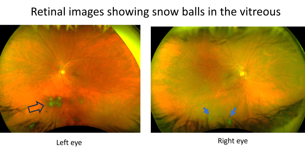
The exam showed normal vision, normal eye pressure, and interestingly no vitreous detachment. However, there were some cells in the vitreous in the left eye (again refer to Figure 1 to see where the vitreous is in the eye). The retina appeared normal without any tear or detachment. A closer looked showed that there were even cells in anterior chamber, the front chamber of the eye (in contrast, the vitreous is the back chamber of the eye). 

Figure 1. The vitreous is a large space filled with a jelly inside the eye. In Edward’s case, this jelly now contains many inflammatory cells causing him to see floaters. Image from National Eye Institute [1].

So this was a form of uveitis, which is less common, but also can cause floaters, which I talked about in a previous case also (link). 

Uveitis is the inflammation of the colored layer of the eye, called uvea (Figure 1). Injuries can cause this inflammation, but so can infections, autoimmune diseases, cancer and sometimes no reason that we can find.

The question is, what is causing the uveitis?

More about this patient

I asked him if there was anything else going on besides the eyes? 

He said, ‘I have had a skin rash on my back before the floaters showed up. My dermatologist gave me a fungal cream but it’s not working and I am going back to see him next week.’

Otherwise he was in his usual health with no complaints.

Looking at his other office visits, I could see that he fought two cancers, once 10 years ago and once 5 years ago. Fortunately both were found in stage 1 and adequately treated.

He was HIV positive, but had normal CD4 values and was taking his anti-virals consistently. He was also being tested regularly on chlamydia and syphilis and negative in these in the last labs 4 months ago.

Based on this history, cancer is a concern. But HIV related etiologies are also possible. These both can cause uveitis. Further, inflammatory causes (non-cancer, non-infectious) are also possible. 

Cause of uveitis revealed by skin test

I promptly referred him to our uveitis specialist, who will run a battery of tests and imaging to figure out what is the cause.

However, it was his dermatologist that nailed the diagnosis and sent him for the definitive treatment.

Since the rash did not improve with medical treatment, his dermatologist performed a skin biopsy, which showed a very small bug (called spirochetes), the causative agent for the rash, and also the uveitis in the eye. These spirochetes are the microorganisms that cause syphilis, a sexually-transmitted disease that is sometimes found to be more prevalent in those with positive HIV. 

In this case, a simple skin biopsy saved him the trouble of a battery of tests and imaging procedures, which was the silver lining of his unfortunate symptoms. The treatment, IV penicillin, is usually quite effective.

Additional thoughts

In the end, this outcome might be the best it can be. Cancer in the eye, such as lymphoma, would be much more difficult to get rid of, not to mention possibility of death.

As an eye doctor, the most important thing is not to mis-diagnose this special form of uveitis as the good old vitreous detachment, even though vitreous degeneration or detachment is the horse, and uveitis is the zebra.

And syphilis, a great mimicker of all forms of uveitis, should always be tested, especially if the patient is at risk.

So if you have new floaters, please reach out to an eye doctor, because your retina may be at risk and it could be something else.

Learn more about uveitis from this reference below.

Reference

[1] Uveitis, National Eye Institute, https://www.nei.nih.gov/learn-about-eye-health/eye-conditions-and-diseases/uveitis

Pink eyes after recovering from a cold?

Juan Ding, OD, PhD

Just the first week in 2024 back in clinic, I started seeing several pink eye cases. Some of these have one thing in common, that the patient recently had covid or other viral illnesses.

Patient #1 had covid related pneumonia and recovered 4 weeks later, her red eyes onset just a few days after pneumonia diagnosis. With treatment of doxycycline respiratory symptoms almost resolved, but red and painful eyes persisted. There was even involvement of the cornea and the iris (keratitis and uveitis).

Patient #2 had covid with bad symptoms (upper respiratory, fever and fatigue), but no pneumonia. Red eyes onset after she almost recovered fully from covid. The conjunctivitis appeared viral, no involvement elsewhere in the eye.

Patient #3 had cold symptoms, but did not test positive for covid, and we don’t know what virus he had. After he almost recovered from the coughing symptoms, his eyes became red. Only conjunctivitis and no cornea involvement.

There were earlier reports in 2020 when covid first came out that pink eye, or conjunctivitis, could be the first symptom and sometimes the only symptom of covid. I have not read much that the eye symptoms come after acute covid symptom resolution. But apparently that could happen, as I saw a cluster of these just in 2 days. It is common to see viral conjunctivitis showing up at the same time or some days after the cold symptoms such as in patient #1, not too often when the cold symptoms completely resolve like in patients #2 and 3. Of course, patients could be having different interpretations about the resolution of the respiratory symptoms- it’s possible they are just getting much better and not completely over the cold.

Anyway, in the peak of all the viral infections in January and February, you may come down with red eyes when you have a cold, sometimes even after coming out of a cold. Sometimes it’s not just a pink eye, it can be infection or inflammation of the deeper structure of the eye, and it is best go see an eye doctor.

Leave a comment below, when you had a pink eye, was it stand alone, or before or after cold symptoms?

Winning lottery twice

Juan Ding, OD, PhD

This is a case where two rare eye conditions happen to the same patient, as if they win the lottery (of the unfortunate) twice.

I saw Mary (not her real name) in her early 50s last year. She came to me because she had seen new floaters in the left eye for 1 week. I have written about floaters previously, you can find them here, here, and here for more information. 

There are floaters that affect almost everyone: the regular floaters and the acute PVD (posterior vitreous detachment) floaters, and there are also weird floaters that affect only a few who win a lottery in the weird eye condition category. Mary won that lottery unfortunately. There were snow balls in her left eye, inside the jelly we call vitreous. You can see these snowballs when looking at her retina (Figure 1).

Figure 1. Initially only the left eye had intermediate uveitis, with snow balls (immune cells aggregates) in the vitreous as shown by the arrow head on the left; months later the right eye also developed intermediate uveitis with snow balls as shown by two arrows on the right.

This is called vitritis, an inflammation of the vitreous (which is the jelly inside the eyeball). When vitritis happens alone without inflammation in other parts of the eye, it is called intermediate uveitis. The most common symptom of intermediate uveitis is floaters. The snow balls are actually the immune cells accumulating inside the vitreous. It is critical we find out why inflammation happens there, because some of them can have serious health consequences to a patient’s vision and even life.

Common causes can be infections such as tuberculosis, leprosy, Lyme’s disease, syphilis, toxocariasis; autoimmune diseases such as sarcoidosis, multiple sclerosis, inflammatory bowel disease, Sjogren’s disease, tubulointerstitial nephritis and uveitis (TINU) syndrome; and cancer such as lymphoma.

I referred Mary to a uveitis specialist, who did an extensive workup and did not find a cause for her intermediate uveitis. This happens more often than not in medicine. But at least the infectious causes had been ruled out, so immune suppression treatment can be started; if the cause is an infection, then immune suppression will make things worse. She was started on oral steroids to treat the presumed autoimmune cause (though unclear what it is. She saw a rheumatologist but no diagnosis was found). She got better, but vitritis returned after the steroid taper. At that time they also found increased transaminase indicating a liver problem so no more oral steroids were given. She saw a GI specialist, who did not find a cause of increased transaminase level, and a few months later the level got better on its own.

Given the potential liver issue, no oral steroids, but eye drop steroids were given to her to treat the vitritis. She got better for sometime, but then her eye pressure spiked as a side effect of the steroid eye drops, which had to be discontinued. Meanwhile vitritis started in her right eye as well (Figure 1). 

Next step of treatment would be a systemic agent that does not cause liver damage. The uveitis specialist was able to get insurance to pay for Humira (Adalimumab), and with this medication, her vitritis and floaters finally got better in both eyes up until now. This medication blocks the activity of TNF, a molecule used by our body’s immune system to create inflammation. Less TNF means less inflammation in the eyes, and it has been working for her.

She came back to me again for new glasses now. A year ago when she first came to see me, she was wearing her new glasses of just 3 months. She had myopia and astigmatism in both eyes, more in the right eye. In that visit 1 year ago I actually found more astigmatism in the right eye and gave her an updated script. It was a little unusual to have a change in just 3 months, but she had other things to worry about for her eyes (the vitritis), so this was not pursued.  Now her vision was blurry, and the right eye had a further increase in astigmatism. This is not normal. So I ordered a corneal topography, and sure enough, she actually has keratoconus in both eyes (Figure 2).

Figure 2. Surface map showing steepening of inferior cornea (evidenced by red color) in both eyes, characteristic of keratoconus.

Keratoconus is not a common eye condition, and you can read about it in my previous articles here and here. It often shows up among young people, as the disease tends to progress at a young age. For her to progress and get a diagnosis at 53, that was unusual. Talking about coincidences, I happened to have another 52 year old female patient on that same day whom I diagnosed keratoconus 6 months ago. She also started having vision problems only in the recent couple of years. Life sometimes cannot be explained by just random events, or we humans are just too good at picking up anomalies. 

Anyway, the prevalence of intermediate uveitis is very low, about 6 out of 100,000 people. The prevalence of keratoconus is higher, about 5%, but still not a common condition. For her to have both, it is a chance of 3 out of 1 million. Winning two lotteries!

I would not want her luck. Getting either one of these conditions is a frustrating journey in gaining vision back. She is frequently in doctor’s offices, taking eye drops and medications probably life-long if she’s lucky enough to have the eye inflammation under control, may need eye surgeries and special contact lenses if vision continues to worsen with keratoconus. 

After the new measurement of her eye powers, I gave her a new prescription. She was happy and said, ‘I haven’t had new glasses for over a year, now I’m ready to get new ones and see better.’ In fact, she sounded more positive than many of my healthy patients whose only problem was needing glasses. Happiness is a state of mind, regardless of the conditions that you may have physically. 

References

[1] https://eyewiki.aao.org/Intermediate_Uveitis

[2] Jacinto Santodomingo-Rubido, et al, Keratoconus: An updated review, Contact Lens and Anterior Eye, Volume 45, Issue 3, 2022, 101559, ISSN 1367-0484, https://doi.org/10.1016/j.clae.2021.101559. (https://www.sciencedirect.com/science/article/pii/S1367048421002058)

Hard to be a woman

Juan Ding, OD, PhD

I received a message from a patient who sounded desperate. She said that her eyeball is changing color, that she has necrotizing scleritis, and her uveitis doctor is not picking up the phone.  

The last time I saw her was a year ago, she was only 22, a young and vibrant age. However her lazy eye was bothering her a lot. She had it since she was a little baby, the right eye went out with poor vision. She had patching therapy to treat lazy eye, but her vision was still 20/100 in that eye at its best. 

Lazy eye is a condition that develops before 8 years of age. From birth to roughly 8 years, kids’ eyes need to receive sufficient visual stimuli to develop their visual pathway leading to normal vision. A lazy eye could be turning out or turning in, or turning up or down, which prevents this eye from getting the proper visual stimuli, leading to abnormal development of the visual part of the brain corresponding to this eye. As an adult, it becomes very difficult to treat lazy eye, as the visual brain becomes less elastic.

Not only does a lazy eye that has an eye turn affect vision in one eye permanently, it also poses a psychological and social challenge for patients. A very noticeable eye turn can cause reduced self esteem and is a problem beyond just the cosmesis. Many adults with an eye turn seek surgery to make their eyes appear straight, even if that does not change vision. When I asked if she was interested in such an option, she very happily said yes. 

So I referred her to a specialist that does such surgeries for adults.

Most of these surgeries are successful, with a happy patient who feels more confident in their daily lives. Complications are uncommon.

The surgery went well for her. However, very unfortunately, a rare complication happened. Necrotizing scleritis is a rare eye condition which can be caused by autoimmune conditions. She has a preexisting autoimmune condition, psoriatic arthritis. With this condition, patients have  joint pain and red scaly rashes on their skin. Her symptoms were in good control prior to the surgery. However, unfortunately the surgery and sutures triggered inflammation of the white lining of the eyeball, the sclera. Remember she has only one good eye with the other seeing poorly from being a lazy eye. Though eyes are straight, she still relies on only the good eye to see and function. The unfortunate part is that the good eye developed the necrotizing scleritis, as eye turn surgery was typically done on both eyes. Scleritis is a rare condition, necrotizing scleritis is even more rare. It is the most severe form of scleritis, with the tissue undergoing necrosis, or death. Patients have excruciating eye pain and reduced vision. The prognosis is poor, with high likelihood of vision loss.

No one could foresee such a complication to happen, but it happened. She was seeing uveitis specialists from the best eye hospital in the area, as well as rheumatologists. The silver lining of this unfortunate event is that after some time, her eye was stabilized and under control with a medication called methotrexate as well as multiple eye drops. She was doing well for some time and her vision was preserved. 

Until she got pregnant. She should not get pregnant while taking the medication that she’s taking for her active autoimmune condition. It was not planned. But she’s happy with her fiance and she’s scared but loves her baby. Methotrexate is a pregnancy category X drug, which means it is not recommended to take during pregnancy. It may cause miscarriage and birth defects of the baby. 

What now? She had to stop the medication. Her eye condition took a turn. It’s getting worse again, and she is being referred from one uveitis specialist to the next. She was not able to get an appointment and she told me that her eyeball is changing color. The color change in scleritis means the sclera tissue is getting thin, to the point the underlying choroid is showing through. 

I am not a uveitis specialist, her case is beyond my specialty. I reached out to her specialists- I do not want to get into the details of the process- and I believe that she will be seen and treated soon.

It is not easy to be a woman. First off, women are more likely to have autoimmune conditions and more likely have severe symptoms. Well she is one of these women. Autoimmune diseases can affect young people, and she is young. Second, most medications for autoimmune conditions are not pregnancy friendly. For women at child-bearing age, if they are battling active autoimmune diseases, they hardly have a choice to have kids. If they do get pregnant, they may have to hold off the treatment and risk their conditions getting worse. There is no winning for them.

For my patient, she has bad luck to incur a rare complication after a usually very safe surgery, a surgery that she rightfully deserves. The pregnancy should have been prevented. But accidents can happen. On the one hand, she is risking losing vision in the only good eye without the medication, and on the other, she would be risking the baby if she were to resume the medication. Difficult choice and I feel very sorry that she has to choose.

If it were you, how would you choose?

What, an eye AND ear problem?

by Juan Ding, OD, PhD

A 70 year old Asian male complained of eye pain and redness in both eyes, in addition, left ear was swollen and mildly painful, and not hearing too well. He was found to have episcleritis (inflammation of a deeper layer in the white part of the eye) in both eyes and iritis (inflammation of the iris) in the left eye. Combination of these symptoms are suspicious of a condition called relapsing polychondritis (RP). This is an autoimmune disease that targets the cartilage tissue of the body, for example the ear, nose, and the trachea. Eye inflammation can also occur. Since it’s autoimmune in nature, the treatment would be anti-inflammatory, using for example corticosteroids and non-steroidal antiinflammatory drugs (NSAIDs). If necessary, some disease modifying agent such as methotrexate can also be used. He was started on naproxen 500 mg twice daily, as well as an NSAID and steroid eye drops, and his symptoms went away. So in the end he did not need to take methotrexate.

He also had a hx of latent tuberculosis (TB). latent TB means a person has been infected with tuberculosis in the past, but does not show any active sign of disease, such as coughing, fever and night sweats. However, latent TB can cause inflammation in any part of the eye, including the episclera and the iris. So in this case, he is also seeing an infectious disease specialist. Since his eye and ear symptoms resolved on NSAIDs and steroids, it was thought that TB was not the cause, but he was treated with isoniazid for 9 months for latent TB without incidence.

Lastly, although RP frequently causes ears to be red and swollen, it does not typically cause hearing loss. His hearing loss was considered to be another cause. He was treated for 1 month after onset of hearing loss (and ear pain) for a viral etiology with valtrex and prednisone, but did not recover hearing.

So the moral of this case is, if they have inflammation of both eyes and ears, don’t just treat the eye, and think a common underlying etiology.

5 years later, patient came back to see me, his eyes are quiet, as his ears. He does report occasional eye sensation reminiscent of past iritis, but always responds well to the prednisolone drops that he’s instructed to use as needed which he used a few times a month. He is currently not taking any medications for RP and doing well. I asked him to come back in 3 months to check eye pressure and check eye inflammation, and that if a flare up happens come right back. Hopefully the relapsing nature of this disease will not recur.

Floaters in a young woman

by Juan Ding, OD, PhD

Walk in patients are fun, especially on a Friday.

27 year old female walked in our open access clinic today, reporting seeing floaters for 1 month in the left eye which bothered her. Otherwise she had no change in vision and no pain. She did not see any flashes. She saw 20/15 each eye and had normal eye pressures. Right eye was completely normal. The left eye had numerous deposits on the back side of the cornea, called KPs, and a few cells floating in the anterior chamber. There were numerous cells in the vitreous. There were two black scars in the retina close to the macula, and a fluffy white lesion with smaller (satellite) white lesions surrounding it. The retinal blood vessels and other parts of the retina, as well as the optic nerve, looked normal (photo below).

toxo

Figure 1. Fundus image showing inactive scars (arrow heads) and an active lesion (arrow).


What is your diagnosis?

On questioning, she admitted to be told to have toxoplasmosis in her left eye in 2015 when she had an eye exam for doing refractive surgery. She was told it was stable before and after her photorefractive keratectomy (PRK) surgery.

Apparently toxoplasmosis has reactivated in her left eye. “Most of the recurrences occur in the second and third decades of life in immunocompetent individuals, and may be triggered by stress or other factors.” according to an American Academy of Ophthalmology article (https://www.aao.org/current-insight/management-of-ocular-toxoplasmosis).

Toxoplasmosis is acquired by eating raw or undercooked meat, vegetables or milk products, or by coming into contact with infected cat litterbox or sandboxes, but contaminated water source has also been linked. Toxoplasmosis can also be congenital, when infection in pregnant women transfer to the fetus.

The treatment for ocular toxoplasmosis can be observation only if the lesion is peripheral and small, as in healthy people this will usually resolve without affecting vision. But if it threatens vision, such as when lesion is close to the macula or optic nerve is involved, or macular edema is present, systemic antibiotics and corticosteroid treatment are usually needed. Topical steroid is also used in the case of anterior uveitis (such as in our patient).

So not all floaters are age-related normal phenomenon.

Drug-induced uveitis or dry eye?

by Juan Ding, OD, PhD

I will try to write about one interesting eye case that I see each day. The purpose is to provide education of eye diseases, vision function and eye care to the public, and maybe to students who study optometry. This is the 2nd of the series.


Woman in her 30s referred by her cancer doctor for red eye. The symptoms were irritation, itchiness, pain, redness, and some crusting, but hardly any photophobia. Cancer doctor informed me that the patient was on two cancer medications, dabrafenib and trametinib, which are known to cause uveitis (inflammation of the middle layer of the eye). Just from the symptoms, it sounded like conjunctivitis or dry eye rather than uveitis. Unexpectedly though, there was indeed inflammation of the iris; the vitreous and retinal were normal, so the inflammation was restricted to the fronter part of the eye, termed iritis. Interestingly, almost all her complaints were actually from the mild dry eye she also had. In contrast, usually iritis would produce a dramatic complaint of pain and photophobia.


My patient had malignant melanoma. She started with chemotherapy with dabrafenib and trametinib for 2 months, followed by surgery, then continued with chemo. About 5 months into the chemo she presented with inflammation of both eyes. Below are the eye side effects of these two medications listed by manufacturer:


“Ocular Toxicities. Retinal vein occlusion (RVO) may lead to macular edema, decreased visual function, neovascularization, and glaucoma. Urgently (within 24 hours) perform ophthalmologic evaluation periodically and at any time for a patient-reported loss of vision or other visual disturbances. Permanently discontinue MEKINIST in patients with documented RVO.

Retinal pigment epithelial detachment (RPED) can occur with MEKINIST administration. Withhold MEKINIST if RPED is diagnosed. If resolution of the RPED is documented on repeat ophthalmologic evaluation within 3 weeks, resume MEKINIST. Reduce the dose or discontinue MEKINIST if no improvement after 3 weeks.

Uveitis (including iritis and iridocyclitis) can occur with TAFINLAR. Perform ophthalmologic evaluation for any visual disturbances. Monitor patients for visual signs and symptoms of uveitis (eg, change in vision, photophobia, and eye pain). Permanently discontinue TAFINLAR for persistent grade 2 or greater uveitis of more than 6 weeks duration.”


Her iritis responded well to prednisolone acetate 1% drops 4 times a day in both eyes, and tapered without incident.


Two months later, she came complaining of bad dry eyes; no uveitis. She got relief with preservative free artificial tears. About the same time, her cancer doctor found sarcoid-like reaction in her lungs.


Another two months later, in a routine follow up eye exam, I found iritis in both of her eyes again, though she had no compliant. It went away again with prednisolone drops.


It is not clear if her two episodes of iritis are due to sarcoidosis, or to drug toxicity; or if her sarcoid is in anyway related to the drugs. It does appear that her symptoms are mostly related to dry eye rather than uveitis. Prior to this she did not have a history of dry eye. Hopefully she can be off of these medications soon and hopefully she will then not suffer from dry eye again.