It was the end of the clinic day. I finished my last patient’s chart and was ready to go, before noticing that a procedure result just popped out. I clicked into it, it’s a visual field testing result of a 60 year old gentleman I saw a few days ago. He complained that after ceiling plaster dropped into his right eye, his right eye could not see in the periphery.
This is a regular patient of mine. When the plaster incident happened 2 weeks ago I was on vacation so he saw another doctor in the practice. He initially went to the emergency room, received plenty of eye washing and antibiotic eye drops. He then saw my colleague two more times and according to the notes, his eye was recovering well. When I saw him his right eye was white and quiet, cornea completely healed, no defect, scar or edema. Internal structures of the eye were also normal. His visual acuity was 20/20. He did have trouble seeing fingers on the right side in his right eye. But his optic nerve appeared healthy. He’s a glaucoma suspect at baseline and I have been monitoring this over 2 years. The RNFL OCT that measures nerve thickness was stable to before. So the question is, why would he have a new visual field defect?
Dr. House says, patients always lie. He insisted that this problem came about after the plaster accident. ‘My eye is a mess’, he said. ‘First it hurt like hell for three whole days, then I cannot see out of my right eye’.
For a chemical injury to hurt the optic nerve, there had to be other signs, like inflammation in the anterior chamber, vitreous and retina. But there was none.
Also it made no sense that he’s losing vision from glaucoma progression, which is typically slow.
It happened suddenly so it’s not a tumor pressing onto his optic nerve either.
Could he be exaggerating because he was frustrated about the whole thing? After all, he waited in the ED for 4 hours, and he was on hold for scheduling to see eye doctors for a long time and did not get call backs.
Of course a formal visual field testing is in order. And that result just arrived in my inbox.
One peek at it, I knew I would not be going home any time soon (Figure 1).
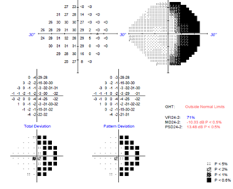

Figure 1. Visual field testing result of the right eye (top image) and the left eye (bottom image). The dark color indicates that the patient was not able to see in that part of the visual field.
The right eye was not seeing the right side of things, just as he complained. Problem is, his left eye was also missing quite a few things on the right side. The left eye was much less severe and it was not picked up in the confrontational visual field test we did in the exam room.
This is what we call a hemianopsia (Figure 1), and it’s an emergency because a stroke was on the differential list.
I called him immediately. I advised him to go to ED immediately, even though he did not have any other stroke symptoms. He does have a history of heart attack and has a pacemaker.
I then called his primary care doctor and she was going to follow up with him.
Hemianopsia happens when one side of the brain that is in charge of vision becomes defective. This can happen with a stroke, a tumor or inflammation. In fact, according to the Cleveland Clinic, 70% of hemianopsia is due to stroke, 15% from brain tumors and 5% from bleeding in the brain [1]. Patients’ eyes can be completely normal, because the problem happens in the brain. It can happen as the only abnormal finding, without other telltale signs of a stroke. The dangerous part is that it is easily missed and over-looked, because the visual acuity can be 20/20, and you don’t find anything wrong with the eyes. Further, patients often describe this in a non-specific manner. Over the years I have heard ‘floaters in the left eye’, ‘my right eye is blurry’, or ‘my eye is a mess’. It is vitally important to always do a confrontational visual field and if suspicious, a formal visual field to clarify and confirm. Otherwise a critical, potentially life-threatening condition may be missed.
Hemianopsia from a stroke may improve over time though may not return to baseline completely, depending on the severity of the damage. Most start recovery within months of the stroke, but it may take up to 18 months for maximum recovery to occur [1]. I will see my patient in 3 months to check his visual field again. To help with vision deficit, certain prism may be used to expand the visual field, but that would the subject of another article.
References:
[1] https://my.clevelandclinic.org/health/diseases/15766-homonymous-hemianopsia-
