Floaters in a young woman

by Juan Ding, OD, PhD

Walk in patients are fun, especially on a Friday.

27 year old female walked in our open access clinic today, reporting seeing floaters for 1 month in the left eye which bothered her. Otherwise she had no change in vision and no pain. She did not see any flashes. She saw 20/15 each eye and had normal eye pressures. Right eye was completely normal. The left eye had numerous deposits on the back side of the cornea, called KPs, and a few cells floating in the anterior chamber. There were numerous cells in the vitreous. There were two black scars in the retina close to the macula, and a fluffy white lesion with smaller (satellite) white lesions surrounding it. The retinal blood vessels and other parts of the retina, as well as the optic nerve, looked normal (photo below).

toxo

Figure 1. Fundus image showing inactive scars (arrow heads) and an active lesion (arrow).


What is your diagnosis?

On questioning, she admitted to be told to have toxoplasmosis in her left eye in 2015 when she had an eye exam for doing refractive surgery. She was told it was stable before and after her photorefractive keratectomy (PRK) surgery.

Apparently toxoplasmosis has reactivated in her left eye. “Most of the recurrences occur in the second and third decades of life in immunocompetent individuals, and may be triggered by stress or other factors.” according to an American Academy of Ophthalmology article (https://www.aao.org/current-insight/management-of-ocular-toxoplasmosis).

Toxoplasmosis is acquired by eating raw or undercooked meat, vegetables or milk products, or by coming into contact with infected cat litterbox or sandboxes, but contaminated water source has also been linked. Toxoplasmosis can also be congenital, when infection in pregnant women transfer to the fetus.

The treatment for ocular toxoplasmosis can be observation only if the lesion is peripheral and small, as in healthy people this will usually resolve without affecting vision. But if it threatens vision, such as when lesion is close to the macula or optic nerve is involved, or macular edema is present, systemic antibiotics and corticosteroid treatment are usually needed. Topical steroid is also used in the case of anterior uveitis (such as in our patient).

So not all floaters are age-related normal phenomenon.

Swollen optic nerves

I am trying to cure my cancer of laziness. So here it goes.

This morning a 26- year old woman came in. Her PCP referred her for eye exam due to headache. She has always suffered from headache since age 8, but recent 5 years it has become worse, sometimes lasting the whole day, and sleeping did not make it better. Initially OTC excedrin helped, but now does not work that well. Her vision is blurry sometimes. She used to wear prescription glasses for reading, but hasn’t for a long time, and now wearing those glasses did not seem to help either. This is an obese female, takes birth control pills, and has a history of type 2 diabetes during pregnancy which resolved since giving birth.

I see a lot of referrals for headache. Almost always the eyes are completely normal. Sure this patient fits the classic “fat fertile female” for idiopathic intracranial hypertension (IIH), but I have seen too many demographics like this that have optic nerves as flat as a shirt. So no expectations here.

However, her optic nerves look slightly elevated. And OCT confirmed thickened retinal nerve fiber layer in some quadrants.

So I guess finally another classical case. I say another because to be fair, this does happen from time to time among the numerous headache patients.


Papilledema due to IIH is often first discovered by eye doctors, because of swollen optic nerves. Patients will then need to go through brain MRI to look for possible mass lesions or blockage of the venous drainage. If none of these, lumbar puncture is done to check for opening pressure of the cerebrospinal fluid (CSF) and look for signs of infection or inflammation in the CSF. If pressure is high but CSF is clear, then a diagnosis of IIH can be made. Patients may be prescribed diamox, which is a carbonic anhydrase inhibitor that reduces CSF pressure; and encouraged to lose weight because for unknown reason, IIH strongly associates with being over-weight. Papilledema usually does not affect vision, but chronic cases may lead to visual field loss and even reduced visual acuity. So we are always on the lookout for optic nerve edema in a patient with headache.

Hallucination day

I will try to write about one interesting eye case that I see each day. (Though I have been guilty of not updating this for a long time, not for lack of cases, but due to my own laziness.) The purpose is to provide education of eye diseases, vision function and eye care to the public, and maybe to students who study optometry.


You know that sometimes unusual cases happen in clusters, like when you see three central serous retinopathy or two posterior polymorphous corneal dystrophy patients on the same day? Obviously if you are a tertiary referral center then that can be common, but for a primary eye care practice, it can be striking. I just saw two patients with visual hallucination today, that’s about the total number of hallucination cases I have ever seen since starting this primary eye care clinic 2 years ago.

68 year old lady stated she was seeing a woman in old fashioned clothes climbing a ladder in her living room, and specifically the woman had very hairy legs. She knew it was a hallucination, and went to ED for it. She was hospitalized for a few days for stroke workup including brain CT and then MRI; saw a cardiologist and then a neurologist. She told me 5 doctors were working on this for her. She had a few systemic issues including diabetes but she was not taking any medications as she stopped all these sometime ago. Anyway it turns out her visual hallucination is most likely a result of Charles Bonnet Syndrome, since her left eye has developed a dense cataract with a visual acuity of 20/80, and this moderate vision loss led to visual release hallucination. She is going to call her cataract surgeon for an evaluation.

At 3 PM a 59 old guy walked in per his cardiologist’s urgent referral. Cardiologist told me it’s a loss of vision post operation, so my immediate thought went to optic neuropathy or central retinal artery occlusion (CRAO) or something along that line. His left eye had a stick penetrating as a child and could never see much since then. Right eye was the good eye. So I assumed that his left eye was not seeing well now. So when he read 20/15 on the eye chart, I almost threw him out- this guys sees better than a 30 year old, why wasting my time? On further questioning, he admitted it’s not that the right eye was not seeing well, rather his left eye started seeing too much that bothered him. This made no sense. It was only in hindsight that I realized why he was being so vague about his symptoms. First, hallucinations are things that do not follow logic or make sense; 2nd, people in general feel embarrassed when talking about their hallucinations for fear they will be thought of as crazy. Eventually I got the story out of him. Last Monday he had a heart surgery and was released home last Friday, when he started noticing seeing colors like through prism glasses and all sorts of shapes like pumpkin and so on. He noticed these in his weak left eye but also in the normal right eye. Only when he closed both eyes did they go away. This was concerning to him because when he talked with people he would see these things on people’s faces and made it hard for him to focus.

You may guess the rest of the eye exam. Yes he had completely normal eye health in the right eye, and left eye was aphakic (having no lens) and had glaucoma but he’s seeing an outside eye doctor every 3 months for this and his eye pressure today was 16 (normal).

Could it be Charles Bonnet after so many years of poor vision in that left eye? Maybe. Was it a coincidence that he started this after a major surgery? Not sure. Since he did take multiple medications (quite a few medications including some hypertension meds, antidepressants, even some antibiotics are associated with visual hallucinations), and it’s after a heart surgery, I referred him to see a neurologist to rule out other causes of hallucinations.

Charles Bonnet Syndrome was first described by Charles Bonnet, a Swiss philosopher, who witnessed his near-blind grandfather seeing things that were not there. These hallucinations happen because of lack of visual input to the brain to suppress certain spontaneous firing of visual cortex neurons. Any form of visual loss may cause it, but commonly conditions such as age-related macular degeneration, glaucoma, dense cataract, etc are associated with Charles Bonnet Syndrome. By the way, with both my patients today visual hallucination was not associated with auditory or other sensory hallucinations, and patients were aware that these things they saw were not real. These were additional criteria for Charles Bonnet Syndrome.

Drug-induced uveitis or dry eye?

by Juan Ding, OD, PhD

I will try to write about one interesting eye case that I see each day. The purpose is to provide education of eye diseases, vision function and eye care to the public, and maybe to students who study optometry. This is the 2nd of the series.


Woman in her 30s referred by her cancer doctor for red eye. The symptoms were irritation, itchiness, pain, redness, and some crusting, but hardly any photophobia. Cancer doctor informed me that the patient was on two cancer medications, dabrafenib and trametinib, which are known to cause uveitis (inflammation of the middle layer of the eye). Just from the symptoms, it sounded like conjunctivitis or dry eye rather than uveitis. Unexpectedly though, there was indeed inflammation of the iris; the vitreous and retinal were normal, so the inflammation was restricted to the fronter part of the eye, termed iritis. Interestingly, almost all her complaints were actually from the mild dry eye she also had. In contrast, usually iritis would produce a dramatic complaint of pain and photophobia.


My patient had malignant melanoma. She started with chemotherapy with dabrafenib and trametinib for 2 months, followed by surgery, then continued with chemo. About 5 months into the chemo she presented with inflammation of both eyes. Below are the eye side effects of these two medications listed by manufacturer:


“Ocular Toxicities. Retinal vein occlusion (RVO) may lead to macular edema, decreased visual function, neovascularization, and glaucoma. Urgently (within 24 hours) perform ophthalmologic evaluation periodically and at any time for a patient-reported loss of vision or other visual disturbances. Permanently discontinue MEKINIST in patients with documented RVO.

Retinal pigment epithelial detachment (RPED) can occur with MEKINIST administration. Withhold MEKINIST if RPED is diagnosed. If resolution of the RPED is documented on repeat ophthalmologic evaluation within 3 weeks, resume MEKINIST. Reduce the dose or discontinue MEKINIST if no improvement after 3 weeks.

Uveitis (including iritis and iridocyclitis) can occur with TAFINLAR. Perform ophthalmologic evaluation for any visual disturbances. Monitor patients for visual signs and symptoms of uveitis (eg, change in vision, photophobia, and eye pain). Permanently discontinue TAFINLAR for persistent grade 2 or greater uveitis of more than 6 weeks duration.”


Her iritis responded well to prednisolone acetate 1% drops 4 times a day in both eyes, and tapered without incident.


Two months later, she came complaining of bad dry eyes; no uveitis. She got relief with preservative free artificial tears. About the same time, her cancer doctor found sarcoid-like reaction in her lungs.


Another two months later, in a routine follow up eye exam, I found iritis in both of her eyes again, though she had no compliant. It went away again with prednisolone drops.


It is not clear if her two episodes of iritis are due to sarcoidosis, or to drug toxicity; or if her sarcoid is in anyway related to the drugs. It does appear that her symptoms are mostly related to dry eye rather than uveitis. Prior to this she did not have a history of dry eye. Hopefully she can be off of these medications soon and hopefully she will then not suffer from dry eye again.

Accommodative insufficiency and convergence insufficiency

by Juan Ding, OD, PhD

I will try to write about one interesting eye case that I see each day. This is the first of the series.

A pretty woman with meticulous makeup (at least for me) in her early 40s complained about difficulty reading and seeing distance. Also had bouts of double vision a few months ago. She has a history of bipolar disorder, hypothyroidism and alcohol use, and goes in and out of rehab for alcohol problem.


This is the second time I see her. I saw her 3 weeks ago for the first time. As in any comprehensive eye exam, when I saw her, she’s already been worked up by the technician with her pupils dilated. Unable to make her see 20/20 with refraction, yet with a complete normal eye exam otherwise, I suspected a functional problem. So here she is again today, sitting in my chair, only able to see 20/60 letters for distance, and 20/100 letters at close up. Looking at her auto-refractor reading, I know right away that she will not respond to phoropter refraction. So I did the good old trial frame. With +0.25 D of astigmatism correction and +0.25 of sphere, she’s seeing the 20/20 line. Makes no sense even for a 70 year old to see so poorly with such a minor prescription. For near, she requires a +2.00 add, which people usually don’t need until 55 years and older.


I then did other tests to find out that her accommodation is indeed non-existent, and the ability of her two eyes to converge and diverge is also minimal.


I tried two pairs of glasses on for her, one for distance and one for near, and she’s comfortably seeing everything clearly. So I gave her the glass prescription, and ask her to come back if her vision is not improved with glasses.


My diagnosis for her is accommodative insufficiency and convergence insufficiency. These are functions of the eyes to focus and to look at objects at the same point, respectively. I have seen quite a few young women with these issues, and many of them with a psychiatric disorder such as bipolar, depression or anxiety. However, the visual functional deficit is not a psychological one, their eyes truly cannot perform, and glasses can usually help.


It has been shown that certain psychiatric medications such as ritalin can lead to accommodative insufficiency which makes convergence insufficiency worse (http://www.pressvision.com/pdf/hong_press.pdf). It is not known whether the medications that these patients are usually on, or their psychological issues, contribute to their accommodative and vergence dysfunction. Maybe both. Maybe another underlying cause results in their psychological and eye issues.


Common symptoms of accommodative insufficiency include:

  • difficult to focus,
  • blurry vision (especially when reading, using computer or phone)
  • eye strain

Common symptoms of convergence insufficiency include

  • double vision (especially when reading, using computer or phone)
  • eye strain.


Treatment for both:

  • Wear glasses: sometimes bifocals or progressive addition lenses are needed, and sometimes prism may help further.
  • Vision therapy: this is a set of individualized eye exercises that an optometrist prescribes and trains with motivated patients.

Dry eye caused by medications

Do you know that among the top 100 best-selling drugs in the US, 22 of them can cause dry eye? In fact, 62% of dry eye cases in the elderly can be attributed to systemic medications, including nonsteroidal anti-inflammatory drugs (NSAIDs), diuretics, vasodilators, analgesics/antipyretics, antiulcer agents, sulfonylureas, cardiac glycosides, anxiolytics/benzodiazepines, anti-infectives, antidepressants/antipsychotics, hypotensive agents, and antihistamines. “TFOS DEWS II iatrogenic report” has summarized research data in this area and compiled a very nice table below.

Table 1.  Systemic medications that contribute to dry eye.


systemic meds

The reason why these drugs cause dry eye is not completely known, but it is thought that many of them have anticholinergic activity, which means they target intentionally or unintentionally a class of proteins on cells, and these proteins are important for the secretion of tear, mucous and lipid.

Another reason why drugs can cause dry eye is that some drugs are secreted and form crystals in the tear, including amiodarone, aspirin, bisphosphonates, chloroquine, ibuprofen and clofazimine.

Eye drops, can they make your eyes dry?

The answer is yes.

We all know that artificial tear eye drops alleviate dry eye, but some other eye drops can actually cause dry eye or make it worse. One such example is glaucoma eye drops. It is estimated that they cause burning sensation and dry eye in up to 47% of patients. The reason is that most glaucoma eye drops contain a preservative called benzalkonium chloride (BAK), which is a known toxin for cells and causes inflammation on the surface of the eye, as we talked about previously (link here). Interestingly, once switched to preservative-free glaucoma eye drops, dry eye sensation reduce to 16%. It is noteworthy that because of the common dry eye issues associated with glaucoma eye drop use, eye doctors often prescribe artificial tears to be used while patients are using glaucoma drops. However, if patients use an artificial tear that contains preservatives such as BAK, their dry eye may be worse.

For people with significant dry eye, it is recommended that preservative-free forms of glaucoma eye drops be used, and preservative-free artificial tears regularly supplemented as well. If you have such issues, ask your eye doctor about the preservative-free versions of glaucoma drops.

Of note, the medicated eye drops that make dry eye worse can also be due to the active medication itself, in addition to the preservatives.

Again, “TFOS DEWS II iatrogenic report” compiled a table of eye drops that may cause dry eye. A great reference to patients and doctors both.

Table 2. Topical eye drops that cause dry eye.

topical meds.jpg

In the end, while we know that a number of systemic medications as well as topical eye drops can cause or make dry eye worse, this is not to say that we should discontinue these medications. However, it is helpful that doctors and patients are aware of the dry eye side effect of certain medications, and take measures to treat dry eye while on them, or switch to different medications if necessary.

When artificial tears just don’t cut it – Other treatments of dry eye

The most common first-line treatment for dry eye is to use artificial tears (ATs), and these are available OTC. I talked about how to select ATs last time. Eye doctors will also recommend warm compress and lid scrub a couple of times a day, which I also talked about previously. These are really effective for majority of dry eye, which is mild to moderate. If you continue to be bothered by dry eye, there are other treatments available. This article will be about the other treatments your eye doctor may recommend or prescribe. Some of these are products you can buy in store or on the web, others have to be done in a doctor’s office. I have also provided additional links to some of the resources at the end of the article.

Rx eye drops for dry eye

Currently there are two FDA-approved medicated eye drops for dry eye disease, Restasis and Xiidra. The active ingredient for Restasis is Cyclosporine Ophthalmic Emulsion 0.05%. It is an immune suppressant that dampens inflammation on the surface of the eye in chronic dry eye. It was approved by FDA in 2003 based on findings that 15% patients showed increased tear production vs 5% patients in the control group. Caveat with Restasis is that it usually takes 2-3 months of regular using to show an effect.

Xiidra is a Lifitegrast 5% ophthalmic solution, approved by FDA in 2016. Lifitegrast blocks T cell functions, reducing inflammation, and improving dry eye signs and symptoms.

Autologous serum

This is a topical treatment using a patient’s own serum. Your blood is drawn, blood cells are removed, leaving the clear liquid portion called serum.  The serum contains numerous growth factors and hormones, which may promote healing in severe dry eye. However, because of laws and regulations related to blood products, as well as lab processing and storage issues, this is not a popular treatment at the moment; but it is effective, and may gain more insurance coverage in the future.

Intense pulsed light

This was initially used in dermatology to treat acne, skin pigmentation, hair removal and rosacea. Studies have shown that it is safe and effective in treating MGD and improving dry eye. The mechanism of action is not well understood, but may be partially due to the thermal heating of the meibum as well as effects on local blood vessels. It is recommended this procedure carried out multiple times over several months in the doctor’s office.

Punctal occlusion

This is a physical way to block the tear drainage thereby conserving the tears on the eye. Tears are naturally drained via upper and lower tear ducts, which join together and drain into the nasal cavity eventually (Figure 1). By applying a plug to the lower and/or upper puncta (Figure 1), tear will no longer drain easily and therefore stay on the eye for longer. This is a quick and easy in office procedure, and there are plugs that are permanent or temporary based on patients’ need. This treatment is most effective for aqueous deficient dry eye, where the water part of the tear is deficient.

punctal plug.jpg

Figure 1. Punctal plug blocking the lower puncta of tear drainage. Image source: http://www.allaboutvision.com/conditions/punctal-plugs.htm


Moisture chamber spectacles

Moisture chamber spectacles are eyeglasses that provide a humid environment and minimizing airflow over the ocular surface. These have been shown to be effective in some case reports.

Tear stimulation

A number of topical eye drops containing medication have been developed in the hope of stimulating tears, some failed clinical trials, others still in development. There is one device that is currently available, called the TrueTear™ Intranasal Tear Neurostimulator (by Allergan), that temporarily stimulates tear production. This product uses a weak electrical stimulation by two probes in the nasal cavities where the nerves are stimulated, resulting in nasal lacrimal reflex (meaning the stimulation of nerves in the nose causes tearing reflex).

Warm compress and its derivatives

Warm compress using a wet towel can be tedious as you have to repeatedly warm it up. There are a variety of microwave heatable products that maintain the temperature for 10 min so no reheating needed.

In addition to these products, a device called LipiFlow® accurately delivers heat to the Meibomian glands to therapeutic levels of 42.5 C. Clinical trials show that the single 12-min procedure of the LipiFlow® system is safe and effective for treating MGD and that the effect can be sustained for 6 months. This treatment is however very expensive and not yet covered by insurance.

Rigid gas permeable scleral lenses

These are hard, large contact lenses worn with saline bathing the entire cornea. Some patients will severe dry eye or other ocular surface diseases find this to be the last resort to alleviate their symptoms. There are a variety of scleral lens designs and brands. One of them is called PROSE (Prosthetic Replacement of the Ocular Surface Ecosystem), which has been around for quite a while and has helped thousands of severe dry eye patients.

One word about new drugs in development

Multiple drugs are in research from various approaches. I have personally been involved in research on hormones that promote Meibomian gland health and thereby have potential to be used as novel therapies to treat dry eye, including growth hormone, insulin-like growth factor-1 and testosterone. In addition, ECF843, a recombinant human protein, lubricin, has recently been licensed by Novartis for up to $ 1 billion. Large scale clinical trials are anticipated.

References and more resources


A patient’s guide to artificial tears

A patient’s guide to artificial tears

The new TFOS DEWS II report provides the latest understanding of dry eye disease (Click here to know more about TFOS DEWS II, the most authoritative literature review worldwide on dry eye disease). How are eye doctors treating dry eye? It turns out, artificial tears are still the first line of treatment. Go to any drug store, and you will find dozens of different bottles of artificial tears. What are in them, what are the differences, and how can you pick out one that is good for you? Let’s take a look. Much of this article is based on information from “TFOS DEWS II Management and Therapy Report”.

DEWS management.png

There are two major types of artificial tears, one that supplements the aqueous, or water part of the tear, and the other that supplements the lipid part of the tear. We know from my previous post (link here) that majority of dry eye is actually caused by Meibomian gland dysfunction (MGD) which results in lipid deficit and rapid evaporation of the tear. Therefore some of the newer artificial tears contain lipid.

Aqueous supplementing artificial tears

To provide lubrication, viscosity enhancing agents are used. These are often referred to as the ‘active ingredient’ in the artificial tears. They include carbomer 940 (polyacrylic acid), carboxymethyl cellulose (CMC), dextran, hyaluronic acid (HA), HP-guar, hydroxypropyl methylcellulose (HPMC), polyvinyl alcohol (PVA), polyvinylpyrrolidone (PVP) and polyethylene glycol. You don’t have to memorize these names at all. I have listed them here in case you want to read the ingredient list on a product and wonder what that weird name means. What matters is that they prolong the time the artificial tear stays on your eye, and that most of these viscosity agents work similarly well.

Some examples: Carboxymethycellulose is used in Refresh and TheraTears; Hydroxypropyl Cellulose is used in Genteal; Hydroxypropyl Guar is used in Systane; and Sodium hyaluronate is used in Blink.

Lipid supplementing artificial tears

These are emulsions where small lipid droplets are suspended in the water base. For example, Systane Balance contains a polar phospholipid, DMPG (dimyristoylphosphatidylglycerol), in addition to Propylene Glycol which is a lubricant; and Retaine MGD contains mineral Oil 0.5% as a lipid.


Gels and ointments

These are thick tears that will stay on the eye for longer, but will cause blurry vision while they stay on the eye, so mostly used at night or before bedtime. They contain high concentration of the viscosity agent, and/or mineral oil. Examples include Genteal Night time gel, Refresh PM and Refresh Lacrilube. These are good choices to use especially if you suffer from dry eyes in the morning or when waking up.


Preservatives vs preservative-free

Preservatives are essential these days to prolong the shelf-life of food, cosmetics, medication, as well as artificial tears. For a ½ oz (15 ml) drop bottle, it will take about a month to finish if you put 4 drops in each eye daily. Of course you will try to avoid touching the tip with finger or eye or anything except for air, and replacing the lid immediately after each use. Still, as soon as you open that sterile bottle, bacteria or fungus free-floating in the air will inevitably get in, let alone the accidental touching of hand, eyelids or other objects. In addition, many people use artificial tears in an as needed manner, this will make each bottle last longer, increasing risk of contamination. A good practice I would recommend is that once you open a bottle, label it with date of opening, and discard after 3 months even if it’s not finished.

So, preservatives allow us to store artificial tears for longer. However, they are known to cause adverse effects on the surface of the eye. The most commonly used preservative in eye drops is called benzalkonium chloride (BAK). “BAK can induce corneal and conjunctival epithelial cell death, damage the corneal nerves, delay corneal wound healing, interfere with tear film stability and cause loss of goblet cells”. To prevent these adverse actions, you should not use preserved artificial tears more than 6 times a day.

Of course, the far better alternative is to use preservative-free (PF) artificial tears. You will know if an artificial tear is PF when you see it is individually packaged in small vials. Once you open one vial, it is good for 24 hours and you should discard it after that. Examples of PF artificial tears include Refresh Celluvisc, TheraTears Liquid Gel, and Retaine MGD.

The problem with these is that they are expensive. A number of new products are now available that use one-way valve dispensers which avoid contamination and allow multiple-use of a PF bottle. I am awaiting for artificial tears to be packaged this way, and hopefully it will be cheaper than the PF tears individually packed. If you know of such a product, let me know by leaving a comment.

Another alternative to the BAK issue is to develop alternative “softer preservatives” which are less harmful to the eye. These are used in eye drops such as glaucoma medications and not just limited to artificial tears. These include oxidative preservatives (sodium chlorite; Purite® and OcuPure™ and sodium perborate; GenAqua™), polyquaternium-1 (Polyquad®) and Sof-Zia™. They cause less adverse reactions on the eye, but still, PF is better, especially if you are using the eye drops multiple times for long-term.


Be careful with redness relief

Some OTC eye drops provide a quick redness relief, such as Visine, ClearEye, Rohto. While they do contain lubricants for dry eye, the redness relief ingredients, which include Tetrahydrozoline or Naphazoline, should not be used long-term. These redness relief ingredients are vasoconstrictors that restrict tiny blood vessels in the eye, making the redness disappear; when used long-term, can actually cause rebound redness, making red eyes worse.

With chronic dry eye, the irritation on the surface of the eye can lead to dilation of the tiny vessels in the conjunctiva, which is the white part of the eye. Therefore the root cause of redness in this case is dry eye itself. Regular use of artificial tears helps to restore normal ocular surface environment, this will get rid of the redness. Forcing the blood vessels to constrict with medication without treating dry eye itself, such as using a vasoconstrictor, will make redness worse if you stop the medication.

In practice, we routinely recommend lubricating artificial tears, but suggest patients to use redness relief only in temporary or emergent situations, such as going to a party in a few hours. In the long term, please control your dry eye, the cause of redness in many cases.

Summary

I have given a brief guide to the various artificial tears available over-the-counter. There are artificial tears that are primarily lubricating via supplementing the water part of the tear, there are also ones that supplement the lipid part. Some are more viscous than others, and the gels and ointments are so thick that they will blur vision for hours, so they are best used before bedtime. Most artificial tears in regular eye drop bottles contain preservatives, which are harmful for the surface of the eye when used in long-term. I would like all patients to use PF artificial tears if possible, but cost is a significant impeding factor.  Finally, watch out for quick redness relief, and do not rely on these long-term, as you will find redness coming back worse once you stop using them. In reality, you may find one particular eye drop to be soothing to you while others are not so great. Don’t be afraid to try a few ones and find the best for you.

A guide to phakic intraocular lenses (pIOLs) for myopia correction

Phakic IOLs are small lenses implanted in the eye to correct refractive error. So far, FDA has approved two lenses to be used for correcting myopia up to -20.00 diopters (D) with astigmatism up to 2.50 D for people 21-45 years of age. These are alternatives to corneal refractive surgery such as LASIK, especially for high myopia, because too much corneal tissue needs to be removed in high myopia in LASIK and it becomes unsafe. As a comparison, LASIK is FDA approved for myopia up to -11.00 D. Click here to read my post on LASIK.

Two implantable lenses approved by FDA

  • The Visian ICL (Implantable Collamer Lens), approved in 2005, is placed behind the iris and in front of the natural lens. It is invisible to the naked eye. The Visian ICL is made of a soft, biocompatible collagen copolymer, and can be folded during implantation, requiring only a small incision of 3 mm.
  • The Verisyse (branded as Artisan in Europe), approved in 2004, is placed in front of the iris, also called an iris-claw lens. The Verisyse lens is made of a rigid plastic, and because it cannot be folded, it requires a larger incision of 6 mm. If you look very carefully, you may see the lens with naked eyes.

Are you a good candidate for pIOL implant?

As mentioned earlier, patients with high myopia outside the range for LASIK can have pIOL implant to correct myopia. However, you still need to meet certain requirements to be a good candidate. Below are some of the important factors to consider.

  1. pIOL is approved for myopia up to -20.00 D, not higher
  2. Two important parameters of the eye need to be determined: the anterior chamber of your eye has to be deep enough; and your corneal endothelium needs to be healthy. This is because the implanted lens needs to have enough room in the eye, otherwise it may block the drainage system of the eye and causes increased eye pressure and glaucoma. In addition, the surgery itself and the lens often cause loss of corneal endothelial cells, as will be discussed further later, therefore a healthy corneal endothelium is also a pre-requisite.
  3. You must have had stable glass prescription for at least a year.
  4. Eye conditions such as cataracts, glaucoma and untreated eye infections will prevent you from being a good candidate.
  5. Systemic contraindications include Sjogren’s syndrome, rheumatoid arthritis, diabetes, HIV and AIDS, and certain medications such as steroids and immunosuppressants may interfere with healing and final outcomes.

What does the surgery involve?

Prior to the surgery, a procedure called laser peripheral iridotomy (LPI) will be performed on the eye that will receive the implant. This procedure makes a hole on the periphery of your iris to prevent the eye pressure from going up during and post-surgery. LPI can be done a week before or on the same day of the surgery. A newer model lens available in Europe and China but not in the US at the moment, Visian ICL V4c, which has a 0.36 mm diameter hole in the center of the lens, allows fluid exchange without the need of this extra procedure.

The surgery itself takes 10 to 30 minutes and is performed on an outpatient basis. Numbing eyes drops are applied, then small incisions made on the cornea to allow the lens to be inserted into the eye. Here is a demonstration video showing the procedure of Visian ICL: https://www.youtube.com/watch?v=wlaAYBefNTo; and for Verisyse lens: https://www.youtube.com/watch?v=Y4GUpMAHA9s.

After the surgery, you will be given antibiotic and anti-inflammatory eye drops to prevent infection and inflammation.

Most patients will notice clear vision shortly after the surgery and their vision stabilizes within a week, but for some patients it may take a few weeks. There is minimum pain although foreign body sensation can be common after the surgery. Most people can resume work and normal daily activities within a few days.

Safety and Efficacy of pIOLs

Phakic IOL implantation is considered to be safe. However, this is still an open-eye surgery, so risks including retinal detachment and endophthalmitis (infection of the entire eye) due to the surgery exist. Generally, the surgery risk is less than that of cataract surgery, but more than LASIK where the eye remains a closed system.

The two types of pIOLs showed equal and comparable safety, predictability, and efficacy [1].

The accuracy of the optics is decent. For Visian ICL, in 41 eyes of 41 patients with myopic refractive errors of -4.00 to -15.25 D, at 8 years, 68.3% and 85.4% of the eyes were within 0.5 and 1.0 D, respectively, of the targeted correction [2].

The vision post-surgery is also good. In FDA trials, the Verisyse have been shown to have 20/40 uncorrected visual acuity (UCVA) or better in 84% of patients after three years and Visian ICLs have been shown to have an UCVA of 20/40 or better in 81% of patients after 3 years [3]. A different study found 60.5% eyes with UCVA 20/20 or better [4].

Considering that this patient population typically have myopia -10.00 D and more, it is not hard to imagine that patient satisfaction is high overall.

So in summary, pIOLs to correct myopia show good safety and efficacy. It may not be completely fair to compare the UCVA post pICLs vs LASIK, as patients in pICLs typically are more myopic than those in LASIK, and we know that some high myopes may not achieve best corrected VA of 20/20 due to myopic retinal changes.

Just by looking at the numbers, the percentage of eyes within targeted correction appears to be lower than that of LASIK (98.6% of eyes reach refraction within 1.00 D of target, and 90.9% of eyes reach within 0.50 D of target refraction). Given that we are comparing two types of refractive surgeries in two different patient populations, one cannot make a conclusion that LASIK achieves better correction than ICL; but as a patient, you can roughly estimate your chance of getting the ideal correction if you are a candidate for LASIK or pIOL.

Complications of ICL

There are several complications, most common are cataract and corneal endothelial cell loss. Verisyse lenses and Visian ICLs are similar, since Visian ICLs are better studied, I will use ICLs as an example for this section. Below are data summarized by a recent meta-analysis [5] unless otherwise indicated.

ICL replacement

The surgery is reversible, meaning that the implant can be taken out or replaced if it is not ideal. Still, no one likes to have a second surgery. Fortunately, only 1.0-2.6% of cases require a replacement of ICL. The common causes for replacement include too long or too short of a distance between the implanted lens and the natural lens.

Cataract

ICL induces a specific cataract called anterior subcapsular cataract (ASC) due to close proximity of the implant to the front part of the natural lens. Based on 8 studies, it is estimated that the incidence of ASC due to ICL implant ranges from 1.1% to 5.9%, and the incidence of ASC cataracts requiring surgery ranges from 0% to 1.8%.

Another review looking at 2592 eyes showed ASC in 5.2% cases, of these, 43.4% were reported within 1 year, 15.4% between 1 and 3 years, and 35.3% ≥ 3 years after ICL implantation [6].

Even though the cataract incidence due to implants remains low, it has been found that for older patients (>40 years of age) and higher myopia (-12.00 D or higher), the risk of cataract were much increased, as high as 28% at 6 year follow up.

High myopia itself is a risk factor for ASC post ICL implant. While clinically significant cataracts occurred in seven (6.6%) of 106 eyes with preoperative myopia of −12.00 D or higher, none occurred in the 420 eyes with preoperative myopia lower than −12.00 D.

Corneal endothelial cell loss

Corneal endothelial cells are important in keeping the cornea clear. These cells do not regenerate and we lose about 0.4% of them every year as a result of normal aging [7]. ICL implantation accelerates the endothelial cell loss to about 7.5% loss at 5 year follow up. Fortunately, this stabilizes after 2-3 years.

Glaucoma

Lens implantation may cause release of pigment from the iris, blocking the drainage system of the eye, thereby increasing eye pressure, leading to glaucoma. Only two  eyes  out  of  526  (0.4%)  were found to have increased  eye pressure requiring  treatment  at  3  years  post op.

Iris atrophy and pupil distortion

Since the surgery involves placing a lens close to the iris at the center where the pupil is, risk for iris atrophy or pupil distortion exist. In following up with 993 eyes undergoing ICL implantation from 1996 to 2008, iris atrophy and pupil abnormality were found in 0.2% of cases [8]. Higher incidence of iris atrophy was found in the iris-claw type Verisyse lens, 11.8% (11 out of 93 eyes) [9]. This may be skewed from being a study with smaller number of patients, but it is also not a surprise, as the Verisyse lens is fixed on the front of the iris.

So, pIOLs or LASIK?

First of all, remember that all surgeries carry risks, and that wearing spectacles is safe and involves no manipulation in your eyes.

However, if refractive surgery is something you want to go for, make sure you know the indications, benefits, risks and complications associated with each. LASIK is approved for myopia up to -11.00 D, and appropriate corneal thickness is required. Common complications include dry eye, and rare but more serious complication such as corneal ectasia can be detrimental to vision.

pIOLs are better for high myopia up to -20.00 D, does not require a thick cornea but requires healthy corneal endothelial cells and a deep anterior chamber. Complications include cataract and corneal endothelial cell loss; and, as a more invasive procedure, retinal detachment and eye infections can happen though very rare. Caution to older patients and those with higher myopia, as risk of cataract goes up significantly in these patient populations.

In terms of efficacy, both are quite good with high patient satisfaction, though LASIK achieving slightly better correction to target.

In terms of long-term safety, LASIK has been approved in the US for over 20 years whereas pIOLs for 12 years, both have been relatively safe with small percentages of complications as discussed in this article and before.

Lastly both procedures are elective and not covered by insurance; pIOL costs about $4,000 per eye, whereas LASIK about $2,000 per eye in the US.

If you are interested in refractive surgeries, make sure you go for complete eye exams and rule out eye and systemic conditions that contraindicate these surgeries. Eye doctors will recommend the appropriate procedure based on your specific eye conditions and systemic health. Make sure you follow doctors’ instructions for pre and post op care to prevent unnecessary complications.


References


  1. Hassaballa, M.A. and T.A. Macky, Phakic intraocular lenses outcomes and complications: Artisan vs Visian ICL. Eye (Lond), 2011. 25(10): p. 1365-70.
  2. Igarashi, A., K. Shimizu, and K. Kamiya, Eight-year follow-up of posterior chamber phakic intraocular lens implantation for moderate to high myopia. American Journal of Ophthalmology, 2014. 157(3): p. 532-9 e1.
  3. Huang, D., et al., Phakic Intraocular Lens Implantation for the Correction of Myopia. Ophthalmology, 2009. 116(11): p. 2244-2258.
  4. Lee, J., et al., Long-term clinical results of posterior chamber phakic intraocular lens implantation to correct myopia. Clin Exp Ophthalmol, 2016. 44(6): p. 481-7.
  5. Packer, M., Meta-analysis and review: effectiveness, safety, and central port design of the intraocular collamer lens. Clin Ophthalmol, 2016. 10: p. 1059-77.
  6. Fernandes, P., et al., Implantable collamer posterior chamber intraocular lenses: a review of potential complications. Journal of Refractive Surgery, 2011. 27(10): p. 765-76.
  7. Galgauskas, S., et al., Age-related changes in corneal thickness and endothelial characteristics. Clinical Interventions in Aging, 2013. 8: p. 1445-1450.
  8. Zhou, T.A., et al., [Mid-long term follow-up results in correction of extreme myopia by posterior chamber phakic intraocular lens]. Zhonghua Yan Ke Za Zhi, 2012. 48(4): p. 307-11.
  9. Benedetti, S., et al., Correction of myopia of 7 to 24 diopters with the Artisan phakic intraocular lens: two-year follow-up. Journal of Refractive Surgery, 2005. 21(2): p. 116-26.


Additional websites that are helpful:

  1. http://www.allaboutvision.com/visionsurgery/implantable-lenses.htm
  2. FDA phakic IOL page: https://www.fda.gov/MedicalDevices/ProductsandMedicalProcedures/ImplantsandProsthetics/PhakicIntraocularLenses/default.htm
  3. https://crstoday.com/articles/2006-apr/crst0406_11-html/


A guide to LASIK, PRK and SMILE

A guide to LASIK, PRK and SMILE

Refractive surgery to correct myopia and hyperopia has been around for many years, with technology evolving from micro blade to all laser today. More and more patients have undergone refractive surgery and said goodbye to their glasses. Many people wish they had done it sooner, but more people are still debating, should I have it done? If so, with all these technologies, which one to choose?

What is the difference between LASIK, PRK and SMILE?

There are several corneal refractive surgeries, the most mature and commonly used of which is LASIK (laser-assisted in situ keratomileusis). FDA approved LASIK to be used in myopia < -11.00 D with astigmatism < 2.00 D; or in hyperopia < +3.00 D with astigmatism < 2.00 D; in patients 18 or older. LASIK operates on cornea, a thin transparent tissue shaped like a convex lens covering the front of the eye. The cornea is composed of five layers, the most front thin layer being the epithelium, followed by a much thicker stromal layer (Figure 1). These two layers are critical for understanding how corneal refractive surgery works.

Picture1

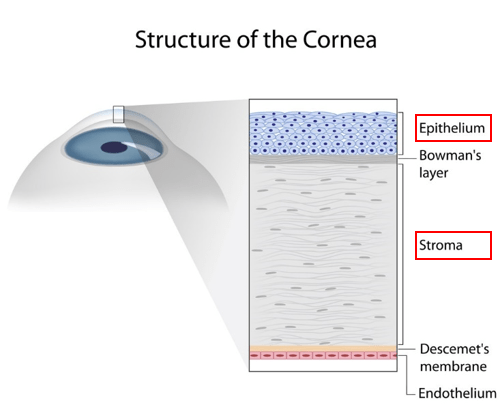
Figure 1. Layers of cornea showing the epithelium and stroma. (Image source: http://discoveryeye.org/treatment-corneal-scratches-and-abrasions/)

Myopia is caused by elongated eye axis resulting in distant light converge in front of the retina (myopia in Figure 2B compared to normal in Figure 2A). You can read more on myopia in my other post [link here]. No surgery can shorten the axis of the eye, but it is surgically possible to reduce the refractive power of the cornea, so that light converges less and reaches the retina again (Figure 2C). In refractive surgery for myopia, a specific amount and shape of the corneal stromal tissue is removed to make the cornea flatter. For hyperopia, opposite strategy is used, the corneal tissue is reconstructed to be steeper.

Picture2.png

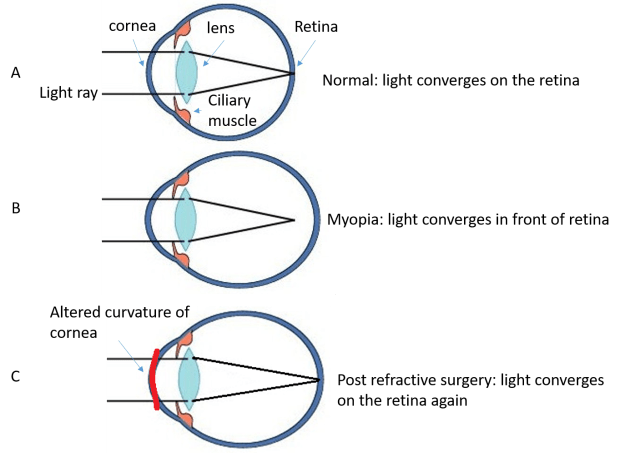
Figure 2:How corneal refractive surgery corrects myopia.


LASIK

The procedure of LASIK is shown in Figure 3. First, a flap is made on the surface (the epithelium and a very thin layer of the stroma) of the cornea (Figure 3A), then a specific amount and shape of the stromal tissue is ablated by laser (Figure 3B), followed by repositioning of the flap (Figure 3C and D). As a result, cornea becomes flatter and myopia is corrected. LASIK technology has become very sophisticated, with the option of wave-front guided customization to minimize higher order aberration, iris registration to track eye movement, and of course, a femtosecond laser used to create the flap. Prior to 2000, the flap was created using a micro blade surgically. Femtosecond laser is a laser that has very high frequency and high energy, which enables quick and accurate incision of tissue. The modern LASIK uses femtosecond laser to create flap, not only being more accurate, but also reducing complications related to the flap compared to micro blade. Still, the flap may dislocate after the surgery, therefore patient is advised not to rub their eyes or do heavy exercises for a week, scuba dive for a month and do kickboxing or jumping out of an airplane for 3 months after LASIK.

Picture3.png

Figure 3:Illustration of LASIK procedure. (Image source: http://www.wfeye.net/type/jzjs/LASIK/)


I remember the first time watching a video on LASIK done on a real patient. I was shocked. It was a huge flap to take off, and it felt painful. Of course it is a painless procedure, it just looks painful. I had mine done 13 years ago in Beijing. Had I watched such a video then, I might not have the gut to go under the table. As research has shown, fear is the number 1 reason to hold back patients in their decision to pursue refractive surgery.

PRK

If a flap on the cornea is not desired due to, for example, being a soldier, an alternative laser surgery called PRK (photorefractive keratectomy) can be used. In this method, the entire corneal epithelium is removed, then the right amount of corneal stromal tissue is ablated similar to that in LASIK. Because no flap is made, complications related to the flap as in LASIK are not an issue. In addition, because the flap contains some stromal tissue, LASIK requires more starting corneal thickness than PRK; thus PRK can be used in certain patients whose cornea thickness is not enough for LASIK. However, the disadvantages of PRK compared to LASIK include more pain and discomfort post operation, as the corneal epithelium which is removed contains large amount of nerves; also, because the epithelium is removed entirely, it takes significantly longer to heal and recover, and extra care is needed to prevent infection. In general, people feel ‘wow, the world is clear’ right after or the next day after LASIK, whereas it will take a week for PRK patients to see the world clearly. You also need to have more frequent follow up visits for PRK vs LASIK. Therefore PRK is usually reserved for those who cannot have LASIK done for various reasons.

SMILE

Now a new technology has come out, Small Incision Lenticule Extraction (SMILE). In this procedure, everything is done by femtosecond laser, and a flap is no longer used. SMILE has been popular in Europe and Asia for a few years now, and has only been approved by FDA recently (September 2016). FDA approved SMILE to be used for myopia of -1.00 to -8.00 with astigmatism of 0.50 D or less, in patients aged 22 and older. In this procedure, a femtosecond laser makes cuts within the cornea, creating a disc-shaped piece of tissue that is removed through a small incision in the surface of the cornea. The small incision is only 2-4 mm compared to a flap diameter of 8-9 mm (or 20 mm circumference). Figure 4 shows the difference in incision on the cornea by SMILE vs LASIK. Because of less damage to the cornea and corneal nerves, it is found that people have better corneal sensitivity and less subjective feeling of irritation and discomfort after SMILE compared to LASIK [1, 2].

Picture4.png

Figure 4:Difference in incision size between SMILE and LASIK (image adapted from: http://www.euroeyes.cn/relex-smile/)


Are they all the same?

In terms of efficacy, all 3 technologies are similar. The difference lies in post op recovery (quick in LASIK and SMILE, slower in PRK), flap related complications (only in LASIK), and dry eye symptoms (least in SMILE). In addition, price is a factor. The latest LASIK technology containing femtosecond laser creating flap, wave-front guided customization, iris registration etc costs about $2,000 per eye (this is only a rough estimate, prices vary in different clinics), with similar price in PRK; while SMILE is still new in the US, the price is about 30% higher than LASIK in China and Singapore.

Should I do it or not?

Refractive surgery is an elective surgery, that is, it is considered cosmetic and not covered by insurance. To do it or not depends on personal needs and value. Not everyone is a suitable candidate for corneal refractive surgery, for example, those with very thin corneas, or myopia >-11.00 D, or those with certain corneal diseases, infections or injuries. Most patients who are good candidates will also have good vision with spectacles or contact lenses. Therefore the motivation of going for refractive surgery really depends on how much you want to do away with glasses or contacts, and whether you are willing to pay.

Many people feel it is inconvenient to wear glasses for work, sports or life in general, or that it is ugly to wear glasses, or that it is inconvenient to wear contact lenses. Lots of people become intolerant to contact lenses due to dry eyes, and this is why they elect to have refractive surgery. One should caution if this is the case. As the biggest complication of LASIK or PRK is dry eye, and those intolerant to contact lenses will find dry eye maybe worse after the surgery even if they no longer need to wear contact lenses.

Some people will calculate the expenses of wearing glasses or contact lenses in a life time and compare that to the surgical expenses. Sometimes though, money is not the most decisive factor. It is interesting how humans are. People who don’t need glasses wear plano spectacles to look fashionable, on the other hand, those with -6.00 D myopia struggle all their lives to be glass free. Human eyes are born to be hyperopic and stay slightly hyperopic through millions of years of evolution (except for a very small percentage of people with congenital eye conditions), but modern civilization and education disrupts the natural environment and the way our eyes are meant to be used (well you are reading this article right now). Read more on myopia progression and treatment here and here. Just like obesity, myopia happened, and medicine struggles to correct it.

What is the success rate of LASIK?

We hear a lot of successful stories; sometimes tragic failures. If you are human, you will be affected by the stories around you. However, let’s also take a look at research and statistics. In evaluating almost 60 thousand eyes with LASIK, it has been found that 99.5% of eyes achieve uncorrected vision of 20/40 or better, 90.8% of eyes achieve uncorrected vision of 20/20 or better; 98.6% of eyes reach refraction within 1.00 D of target, and 90.9% of eyes reach within 0.50 D of target refraction; in addition, only 1.2% of all patients are dissatisfied with the outcome [3]. It is not surprising people are paying a lot of money to have this elective surgery. Of course, if your goal is 20/20 vision, there will be a 10% chance that you will not get it. So be prepared.

Is there any risk to refractive surgeries? What are the complications?

In the case of LASIK, it takes about 5 min to complete the procedure in one eye. With topical anesthetic eye drop, it is a painless procedure. The entire process is controlled by computers and rarely does operation mess up.

As mentioned earlier, the most common complication is dry eye, with 20-55% of patients feeling dry, irritated or burning within 6 months of surgery [4]. This is due to injury of corneal nerves during flap incision and laser ablation of the stromal tissue. Some of the nerves will regenerate, but not necessarily recover to the original state. Fortunately most of the dry eye symptoms can be alleviated by regular use of artificial tears.

Other complications are less common, including flap-related problems (flap dislocation, flap wrinkling, ingrowth of epithelium), ocular surface infection and inflammation. These can be corrected by regular follow up visits, or prevented and treated with proper use of antibiotic and/or steroid eye drops.

One rare but dreaded complication is corneal ectasia, which is the bulging and thinning of the cornea after refractive surgery. It happens in only 0.04% to 0.6% of the cases [5], but once it happens, it will be a nightmare for the patient. I once fitted a young woman with rigid contact lens, who had a history of LASIK for -9.00 myopia. She enjoyed 20/20 vision for 1 week, but then everything changed. Corneal ectasia continued progressing, until it finally stabilized at -16.00 myopia with 3 D of irregular astigmatism. Her vision could not be corrected with spectacle lenses or soft contact lenses, and only by wearing rigid contact lenses could she see well for daily functions.

Some of the factors that increase the risk of corneal ectasia post op include [6]:

  1. The amount of corneal stromal tissue that needs to be removed. The more tissue removed, the more risk for ectasia.
  2. High myopia- obviously higher the myopia, more tissue needs to be removed, and higher the risk.
  3. Thickness of the remaining corneal tissue- thinner the remaining tissue, higher the risk.
  4. Corneal thickness – thin corneas are at higher risk.
  5. Age- younger patients are more prone to corneal ectasia.

In a nutshell, LASIK cuts away a piece of the corneal tissue, the thicker that tissue is, the higher the risk. In some situations where patients have undiagnosed corneal diseases such as keratoconus, their cornea will progress to thinning and bulging quickly after an insult like LASIK. Therefore it is important to have a complete eye exam pre op, including measuring corneal curvature, thickness and corneal topography, and rule out any corneal diseases.

Recently a technique has been developed to conduct corneal cross-linking in combination with LASIK. Cross-linking is known to increase the mechanical strength of the cornea, and has been used successfully to halt the progression of corneal ectasia in certain corneal diseases such as keratoconus. Among 673 eyes in a study, no corneal ectasia occurred with the combo of LASIK and cross-linking, and less regression found compared to LASIK alone [7]. However, this is currently in clinical research and not used in clinic yet.

Will myopia regress after the surgery?

The stroma of human cornea does not regenerate, once it’s removed, it’s gone forever. This is exactly why LASIK works. However, myopia regression can happen in several situations. 1) The patient is still having myopia progression, that is, their axial length is still growing. This is why LASIK can only be done when prescription is stable. FDA approved LASIK to be done in patients 18 years or older. Even though prescription stabilizes after 18 years of age, it is possible for adults up to 40 years of age to have slow myopia progression if they have prolonged near work. 2) Regression can happen if cornea ectasia occur, and not just frank post-op ectasia, sometimes small amount of corneal changes can happen without being clinically significant or being diagnosed. Those with high myopia or thin corneas may slowly progress another 0.50 or 1.00 D of myopia due to small corneal changes.

Will you be glass free forever after the surgery?

Assume the surgery is successful, free of complications or regression, does that mean you will never need glasses again? The answer is that you may not need glasses to see far away, but you will need reading glasses once you hit presbyopic age (about 40). The current technology only corrects for one refraction per eye- if you choose to see distance clearly, you will need to accommodate to see near. When we get older, our lens becomes more rigid, and won’t be able to change power as we look at near. Maybe in future, LASIK technology allows multiple refractions done on the same eye, then you will not need glasses ever. This is not a dream, as multifocal contact lenses are widely used today and working very well.

Summary

  1. It is really a personal preference whether to do refractive surgery or not. It is expensive and not covered by insurance.
  2. If you really want to do it, make sure you go to the eye doctor to have a complete eye exam done and rule out any conditions that contra-indicate refractive surgeries.
  3. There are a number of corneal refractive surgery techniques, the surgeon will help you choose one that is the most suitable for you based on your eye, your systemic health, your work and your lifestyle.
  4. 90% people see 20/20, and 98.8% people are happy with the outcome, but you need to know the potential complications, and be informed and prepared.


Do you have any questions, comments or stories to tell? Leave a comment!

References:

  1. Shen, Z., et al., Dry Eye after Small Incision Lenticule Extraction (SMILE) versus Femtosecond Laser-Assisted in Situ Keratomileusis (FS-LASIK) for Myopia: A Meta-Analysis. PLoS One, 2016. 11(12): p. e0168081.
  2. Zhang, Y., et al., Clinical Outcomes of SMILE and FS-LASIK Used to Treat Myopia: A Meta-analysis. Journal of Refractive Surgery, 2016. 32(4): p. 256-65.
  3. Sandoval, H.P., et al., Modern laser in situ keratomileusis outcomes. Journal of Cataract and Refractive Surgery, 2016. 42(8): p. 1224-34.
  4. Levitt, A.E., et al., Chronic dry eye symptoms after LASIK: parallels and lessons to be learned from other persistent post-operative pain disorders. Mol Pain, 2015. 11: p. 21.
  5. Randleman, J.B., Evaluating risk factors for ectasia: what is the goal of assessing risk? Journal of Refractive Surgery, 2010. 26(4): p. 236-7.
  6. Santhiago, M.R., et al., Ectasia risk factors in refractive surgery. Clin Ophthalmol, 2016. 10: p. 713-20.
  7. Tomita, M., Combined laser in-situ keratomileusis and accelerated corneal cross-linking: an update. Current Opinion in Ophthalmology, 2016. 27(4): p. 304-10.